Log in    RU  


Main page :: Forum :: Ultrasound cases - Sonograms, cine-loops etc. ::

Combined CHD


Tags: Cardiac sonography(Echocardiography), GE Logiq 400 MD, Images, Video, Clinical report, Pediatric

   Posts
 
  Combined CHD
#1

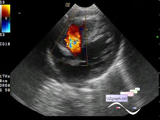
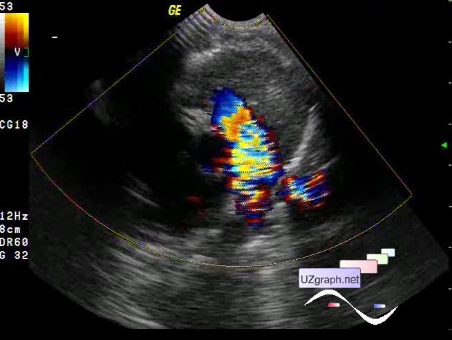
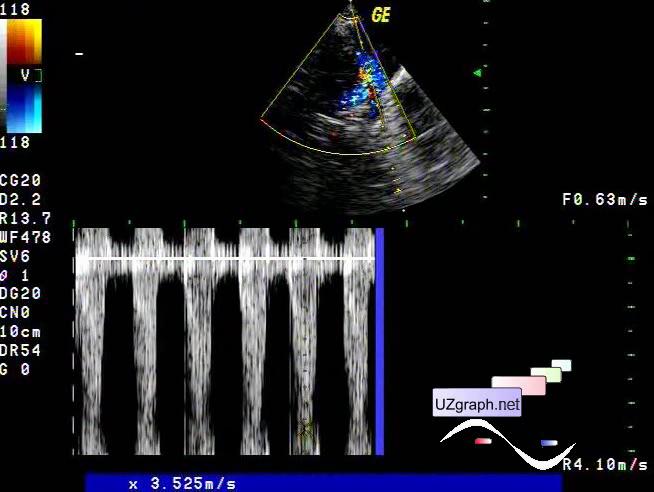
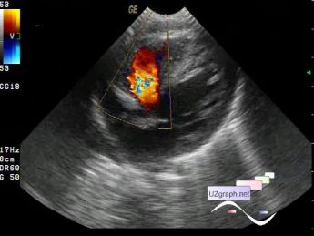
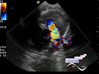
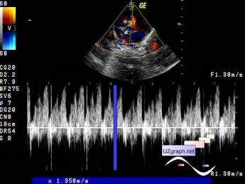
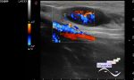
    An infant with a combined congenital heart disease (CHD) on a follow-up echocardiography in a public clinic: perimembranous VSD (ventricular septal defect) (diameter 8 mm) and stenosis of the PA (pulmonary artery) valve (Vmax = 3.5 m/sec), Qp/Qs = 2.5.

    external link

:: attachments(5) ::

:: file 1 ::

:: file 2 ::

:: file 3 ::

:: file 4 ::

:: file 5 ::


 Random 
Ultrasound cases - Linea alba hernia plus
    

Ultrasound cases - Extra flow in the heart
    

Ultrasound cases - Combination of left shoulder lesions
    

Ultrasound cases - Liver cirrhosis and thickened omentum
    

Ultrasound cases - Thyroid lesions 2
    

 Dictionary 
CFM

DPD

IVSd

Color M-mode

PD

PASP

ESV

Qp-Qs

PG

SF


 Calculators 
Teicholz systolic cardiac function

Myocardial mass

Thyroid volume

Type of an infant hip joint

Estimated fetal weight by Ultrasound


 Similar topics 
Ultrasound cases - Floating IAS aneurysm
    

Ultrasound cases - Aortic Stenosis
    

Ultrasound cases - Floating Accessory Pulmonary Valve Tissue
    

Ultrasound cases - Hyperechoic linear structure in the left atrium
    

Ultrasound cases - Hydropericardium / tamponade
    

Ultrasound cases - Coronary fistula
    

Ultrasound cases - Interatrial Communication
    

Ultrasound cases - Tetralogy of Fallot
    

Ultrasound cases - Large Ventricular Septal Defect (VSD)
    

Ultrasound cases - Coronary fistula from the left coronary artery