Log in    RU  


Main page :: Forum :: Ultrasound cases - Sonograms, cine-loops etc. ::

Tetralogy of Fallot postoperative


Tags: Cardiac sonography(Echocardiography), Esaote MyLab 70, Images, Video, Clinical report, Pediatric

   Posts
 
  Tetralogy of Fallot postoperative
#1

    Same child https://m.en.uzgraph.ru/forum/... after operative intervention under supervision in cardiocenter. From last document from cardiocenter: ligation of Blalock anastomosis, radical correction of congenital heart disease with plastic of left ventricle outflow tract(LVOT), PA, patent foramen ovale.

    Comes to me because of long queue to echocardiography in cardiocenter.

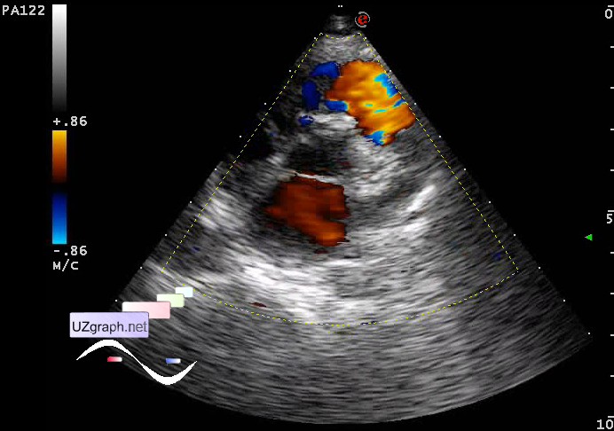
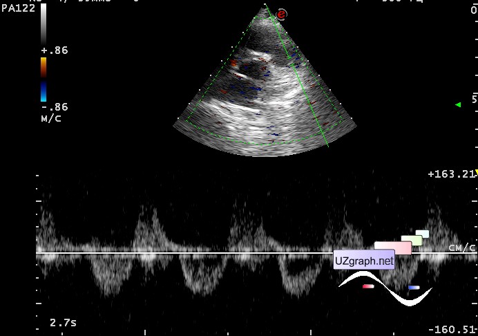
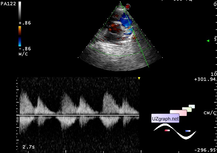
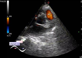
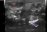
    At echocardiography: at PA bifurcation area retrograde flow(Blalock?); at the area of LVOT flow like IVSD or aortic regurgitation; tricuspidal regurgitation area about 40% of right atrium, PA systolic pressure = 19 mm Hg, PGmax(pressure gradient)PA/RV = 25 mm Hg, AT(acceleration time)PA = 94 msec; EFs(ejection fraction Simpson' s method) = 63%, Qp/Qs=3:1, RVW = 6 mm.

:: attachments(5) ::

:: file 1 ::

:: file 2 ::

:: file 3 ::

:: file 4 ::

:: file 5 ::

  21:56 24-05-2015
  Closed anastomosis?
#2

    Another case...

    Child 2 yo aimed at re-echocardiography, condition after surgery (multi-stage) about Tetralogy of Fallot (in particular was imposed pulmonary-systemic anastomosis, which then, according to the words of accompanying person, was seemingly closed (?). The reason for the re-ultrasound was the previous US, where the sonologist wrote, by the accompanying person words, "strange" information - functioning anastomosis - which, as already mentioned above, seems was closed.

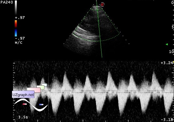
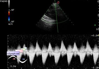
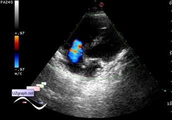
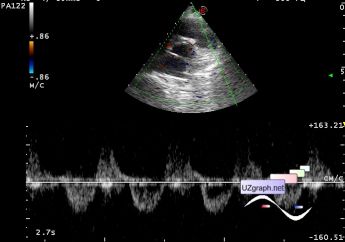
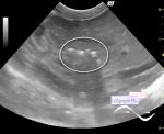
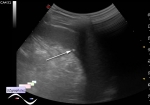
    At current US on CFM in the projection of PA was visualized retrograde flow, in PW just diastolic, in CW systolic-diastolic (2 peaks)

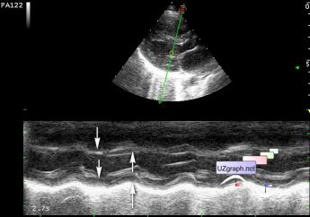
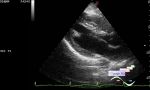
    In M-mode there was a paradoxical movement of the interventricular septum, it moves symmetrically with left ventricle posterior wall.

:: attachments(6) ::

:: file 1 ::

:: file 2 ::

:: file 3 ::

:: file 4 ::

:: file 5 ::

:: file 6 ::


 Random 
Ultrasound cases - Thyroid lesions 2
    

Ultrasound cases - Discharge from the navel
    

Ultrasound cases - Gallbladder polyp simulation
    

Ultrasound cases - Spleen and LN
    

Ultrasound cases - Something on the neck
    

 Dictionary 
GA

FVI

Aliasing doppler artifact

LVEDV

M-mode

EDD

CRL

IVC

LVPWd

MC


 Calculators 
TI-RADS

Teicholz systolic cardiac function

Pulmonary artery pressure

Estimated fetal weight by Ultrasound

Body surface area


 Similar topics 
Ultrasound cases - Bicuspid aortic valve 2
    

Ultrasound cases - Mitral valve cyst
    

Ultrasound cases - Electrode in the right heart after installing of heart pacemaker
    

Ultrasound cases - Chylopericardium, lung atelectasis, cardiomegaly, ascites
    

Ultrasound cases - Asymmetric LVH, aortic regurgitation
    

Ultrasound cases - Coronary fistula from LCA?
    

Ultrasound cases - Floating IAS aneurysm
    

Ultrasound cases - Aortic Stenosis
    

Ultrasound cases - Floating Accessory Pulmonary Valve Tissue
    

Ultrasound cases - Hyperechoic linear structure in the left atrium