Log in    RU  


Main page :: Forum :: Ultrasound cases - Sonograms, cine-loops etc. ::

Appendicular infiltrate


Tags: Abdomen sonography, Gastrointestinal sonography, Images, Video, Clinical report, Esaote MyLab 70, Pediatric

   Posts
 
  Appendicular infiltrate
#1

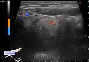
    6 years old girl with clinically suspected acute appendicitis coming to abdominal ultrasound.

    In the right iliac area and pelvis visualized hyperechoic mass with increased bloodflow at DPD, with hypoechoic tubular structure in the middle - possible ultrasound picture of local appendicular infiltrate(complicated acute appendicitis) encapsuled by inflamed omentum(omentitis)

    external link

:: attachments(4) ::

:: file 1 ::

:: file 2 ::

:: file 3 ::

:: file 4 ::


 Random 
Ultrasound cases - Testicular hypoplasia, torsion of cord
    

Ultrasound cases - Partial tears of the lateral meniscus
    

Scientific - Playlist Bones ultrasound

Ultrasound cases - Iliac kidney dystopia of abnormal shape
    

Ultrasound cases - Thickening of the duodenum wall
    

 Dictionary 
PADP

EDD

HC

Color M-mode

MC

PSV

IVC

EFW

Anatomical M-mode

CW


 Calculators 
Pressure gradient

TI-RADS

Body surface area

Gestational age by Ultrasound

Myocardial mass


 Similar topics 
Ultrasound cases - Hirschsprung disease on ultrasound
    

Ultrasound cases - Intestinal Infiltrate
    

Ultrasound cases - Abdominal cryptorchidism
    

Ultrasound cases - Diffuse heterogeneous liver
    

Ultrasound cases - Postoperative abdominal wall fistula
    

Ultrasound cases - Stump appendicitis
    

Ultrasound cases - Liver scar after resection
    

Ultrasound cases - Subdiaphragmatic movable fluid collection (UFO)
    

Ultrasound cases - Lesion of the spleen or the case when it is easy to not notice
    

Ultrasound cases - I’m normal but others ...