Log in    RU  


Main page :: Forum :: Ultrasound cases - Sonograms, cine-loops etc. ::

Foot pain


Tags: Soft tissues sonography, Esaote MyLab 70, Images, Video, Clinical report, Pediatric

   Posts
 
  Foot pain
#1

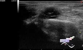
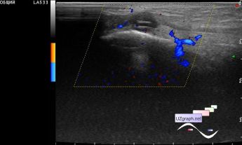
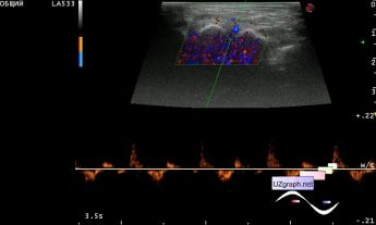
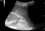
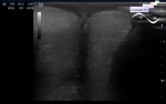
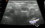
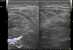
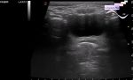
    The teenager was admitted to the emergency department of the Children's City Clinical Hospital with a clinical suspicion of foot phlebitis, complaints of pain in the middle third of the dorsum of the foot, and was sent for an ultrasound scan of the corresponding area.

    On ultrasound in the projection of the pain area, a round lesion of a heterogeneous structure, predominantly an / hypoechoic with hyperechoic linear structures (differential diagnosis: hygroma / ganglion cyst, a site of altered muscle, etc.), with a pronounced increase in blood flow on the DPD, in spectral Doppler mode presumably venous bloodflow.

    external link

:: attachments(4) ::

:: file 1 ::

:: file 2 ::

:: file 3 ::

:: file 4 ::

  23:13 02-08-2021
 
#2

    Some interesting links:

    external link

    external link


 Random 
Ultrasound cases - Hydrocele and rete
    

Ultrasound cases - Thyroid lesion 4
    

Ultrasound cases - Inguinal-scrotal hernia in infant
    

Ultrasound cases - Return of spermatocele
    

Ultrasound cases - The narrowing of the aorta
    

 Dictionary 
LVOT

PRF

Qp-Qs

M-mode

CRL

LVPWd

RVOT

Aliasing doppler artifact

ESV

Ved


 Calculators 
Qp:Qs

Body surface area

Myocardial mass

Resistive index

Type of an infant hip joint


 Similar topics 
Ultrasound cases - Penetrating injury of the femur
    

Ultrasound cases - Cephalohematoma
    

Ultrasound cases - Urachus fistula
    

Ultrasound cases - Cranial mass
    

Ultrasound cases - Pilonidal coccyx cyst
    

Ultrasound cases - Palm fistula, in search of pencil lead
    

Ultrasound cases - Postoperative abdominal wall fistula
    

Ultrasound cases - Nucks cyst
    

Ultrasound cases - Paraproctitis
    

Ultrasound cases - Submandibular area lesion