Log in    RU  


Main page :: Forum :: Ultrasound cases - Sonograms, cine-loops etc. ::

Clavicle area mass


Tags: Soft tissues sonography, Images, Video, Clinical report, Esaote MyLab 70, Pediatric

   Posts
 
  Clavicle area mass
#1

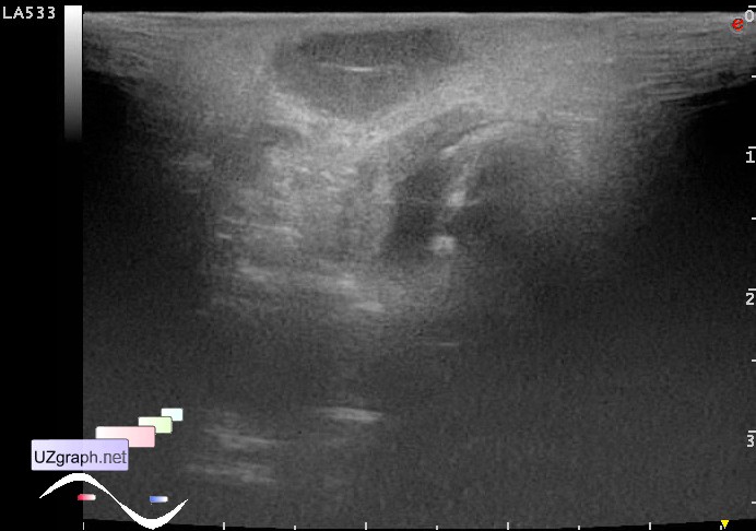
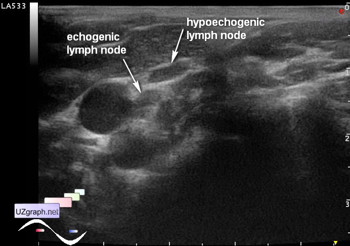
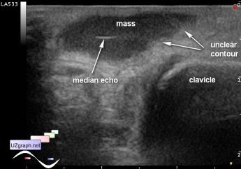
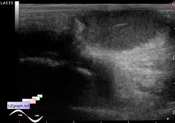
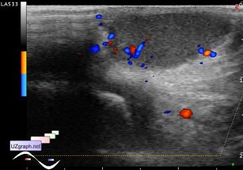
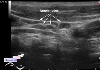
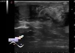
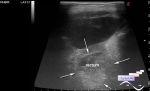
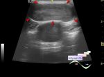
    Child 5 years old came to emergency surgical department with a complaint of mass in the medial edge of the left clavicle, aimed to ultrasound.

    From the words of a parent the mass observed from birth, previously had surgical operation, but there is no documentary evidence of this, at this moment mass arises again, clinically suspected blockage of the sebaceous gland (atheroma/sebaceous cyst).

    Visually mass about 1-2 cm in size, red color with small blue spot.

    At grayscale ultrasound mass with unclear contour, echogenic structure, median echo (?). At DPD with bloodflow inside. Near the sterno-clavicular-mastoid muscle at the same side of body the group of enlarged lymph nodes (in the other side non of the lymph nodes was visualized) of different echogenicity (?).

    Consultation of oncologist was recommended.

    external link

:: attachments(7) ::

:: file 1 ::

:: file 2 ::

:: file 3 ::

:: file 4 ::

:: file 5 ::

:: file 6 ::

:: file 7 ::


 Random 
Ultrasound cases - Onion rings sign
    

Ultrasound cases - Fistula of the lateral surface of the neck in infant
    

Ultrasound cases - Aortic regurgitation
    

Ultrasound cases - Intracranial hemorrhage, CHD
    

Ultrasound cases - Daughters-mothers
    

 Dictionary 
EDD

PW

LVOT

PASP

EDV

IVSd

TV

LVEDV

PADP

LMP


 Calculators 
Thyroid volume

Pressure gradient

Volume (ellipse formula)

Teicholz systolic cardiac function

Pulmonary artery pressure


 Similar topics 
Ultrasound cases - Penetrating injury of the femur
    

Ultrasound cases - Cephalohematoma
    

Ultrasound cases - Urachus fistula
    

Ultrasound cases - Cranial mass
    

Ultrasound cases - Pilonidal coccyx cyst
    

Ultrasound cases - Palm fistula, in search of pencil lead
    

Ultrasound cases - Postoperative abdominal wall fistula
    

Ultrasound cases - Nucks cyst
    

Ultrasound cases - Paraproctitis
    

Ultrasound cases - Submandibular area lesion