Log in    RU  


Main page :: Forum :: Ultrasound cases - Sonograms, cine-loops etc. ::

Double inferior vena cava


Tags: Vascular sonography, Images, Video, Clinical report, GE Logiq 400 MD, Pediatric

   Posts
 
  Double inferior vena cava
#1

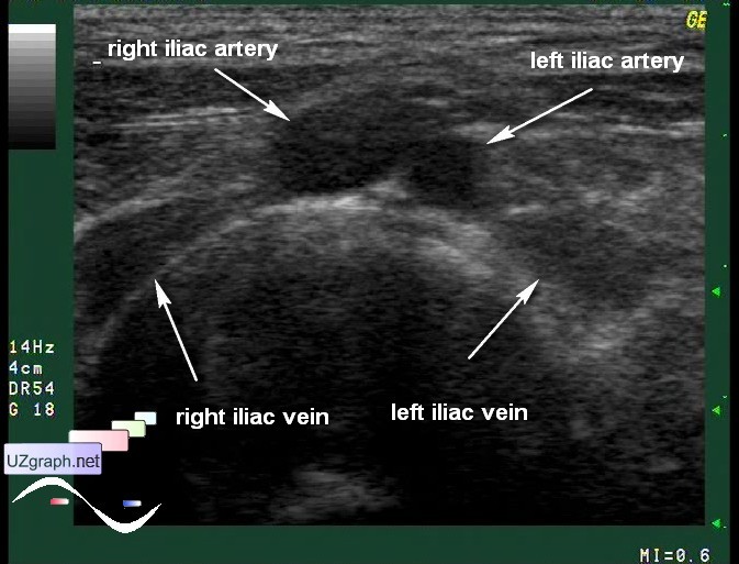
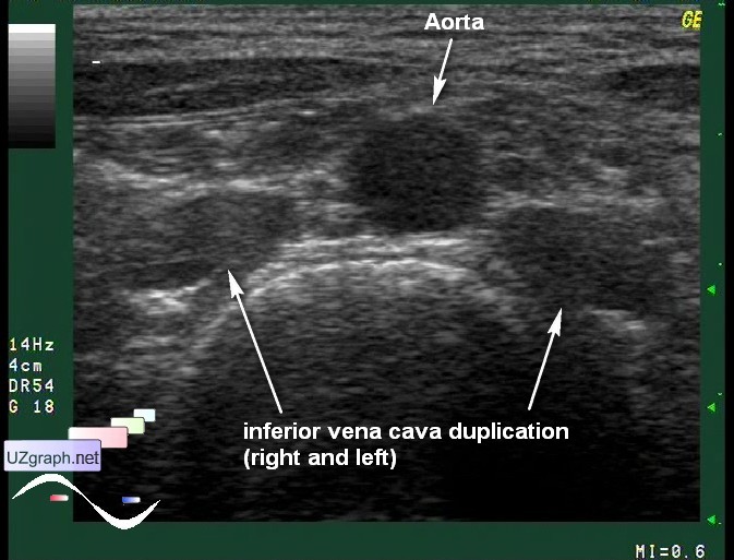
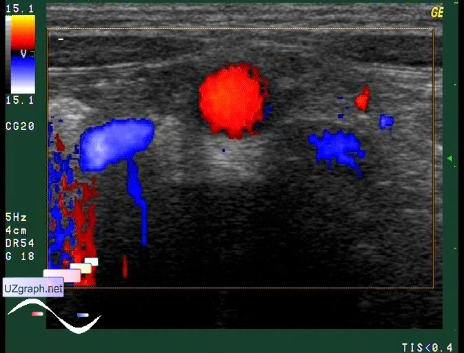
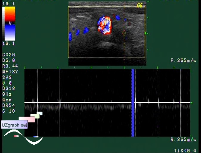
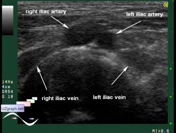
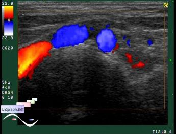
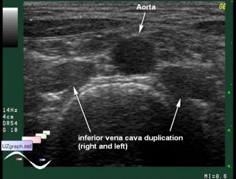
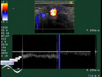
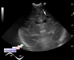
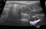
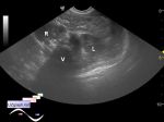
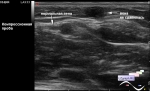
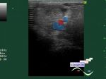
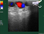
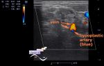
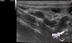
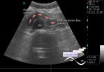
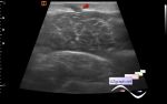
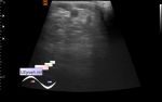
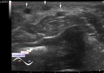
    Teenager cooming for echocardiography. Just in case placed a probe in the low abdomen and found atypical 3 vessels view - aorta and 2 inferior cava veins: right and left.

    It' s a rare congenital anomaly with second left inferior vena cava(LIVC) which drains into the left renal vein. This information should be taking into account if IVC filter placement needed.

    external link

:: attachments(7) ::

:: file 1 ::

:: file 2 ::

:: file 3 ::

:: file 4 ::

:: file 5 ::

:: file 6 ::

:: file 7 ::


 Random 
Ultrasound cases - Tetralogy of Fallot before surgery 2
    

Ultrasound cases - Chord of the brain lateral ventricle
    

Ultrasound cases - Indirect inguinal hernia in a girl
    

Ultrasound cases - Cystic-solid volume of the thyroid gland
    

Ultrasound cases - Wrist edema
    

 Ad 


 Dictionary 
SF

FNA

PADP

LVOT

Doppler angle

HR

LN

Qp-Qs

TI-RADS

LVESV


 Calculators 
Game: Sonographer certification

Area-length (Simpson) formula ejection fraction

Myocardial mass

Teicholz formula ejection fraction

Thyroid volume


 Similar topics 
Ultrasound cases - Subcutaneous abnormal body vessel
    

Ultrasound cases - Complication of a blood test from a vein
    

Ultrasound cases - Anomaly (doubling) of the popliteal vein
    

Ultrasound cases - Varicose disease
    

Ultrasound cases - Vertebral artery hypoplasia in adolescents
    

Ultrasound cases - RLQ phlebectasia
    

Ultrasound cases - Anomaly of caudal aorta
    

Ultrasound cases - Varicose and Baker's cyst
    

Ultrasound cases - Varicose veins of LE
    

Ultrasound cases - Cubital vein thrombosis