Log in    RU  


Main page :: Forum :: Ultrasound cases - Sonograms, cine-loops etc. ::

Hydropericardium / tamponade


Tags: Cardiac sonography(Echocardiography), GE Logiq E, Clinical report, Pediatric

   Posts
 
  Hydropericardium / tamponade
#1

    A teenager with pectus excavatum was sent to the public clinic for follow-up echocardiography due to previously identified mitral valve prolapse with regurgitation of 1 st, with complaints of rapid fatigue during physical exertion.

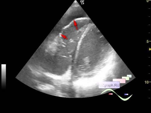
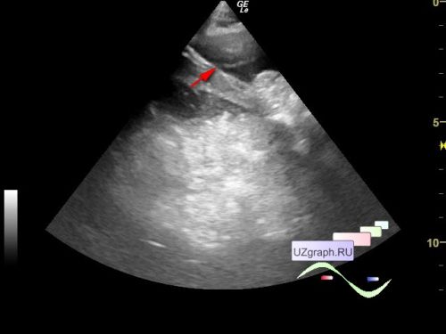
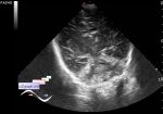
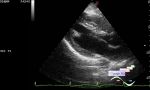
    On the current ultrasound of the heart, prolapse and regurgitation were not detected, but in the projection of the apex of the heart, free fluid is visualized, presumably in the pericardial cavity, a layer of free fluid in different views up to about 1.5 cm.

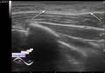
    Inferior vena cava 16 mm, collapses during breathing less than 50%.

    An urgent consultation with a cardiologist was recommended.

    For more see external link

    PS. At the end of the video, we can see the collapse of the right atrium during systole.


 Random 
Ultrasound cases - Cryptorchidism, microlithiasis, bladder lesion
    

Ultrasound cases - Floating IAS aneurysm
    

Ultrasound cases - Sometimes they bite
    

Ultrasound cases - Handle of the LV
    

Ultrasound cases - Sinus venosus ASD / PAPVC
    

 Dictionary 
Gain

TI-RADS

EDV

ESV

PRF

BPD

M-mode

TV

CRL

Anatomical M-mode


 Calculators 
Volume (ellipse formula)

Estimated fetal weight by Ultrasound

Pulmonary artery pressure

Myocardial mass

Teicholz systolic cardiac function


 Similar topics 
Ultrasound cases - Bicuspid aortic valve 2
    

Ultrasound cases - Mitral valve cyst
    

Ultrasound cases - Electrode in the right heart after installing of heart pacemaker
    

Ultrasound cases - Chylopericardium, lung atelectasis, cardiomegaly, ascites
    

Ultrasound cases - Asymmetric LVH, aortic regurgitation
    

Ultrasound cases - Coronary fistula from LCA?
    

Ultrasound cases - Floating IAS aneurysm
    

Ultrasound cases - Aortic Stenosis
    

Ultrasound cases - Floating Accessory Pulmonary Valve Tissue
    

Ultrasound cases - Hyperechoic linear structure in the left atrium