Log in    RU  


Main page :: Forum :: Ultrasound cases - Sonograms, cine-loops etc. ::

Condition after hemangioma cryoablation


Tags: Soft tissues sonography, Images, Video, Clinical report, Esaote MyLab 70, Pediatric

   Posts
 
  Condition after hemangioma cryoabla...
#1

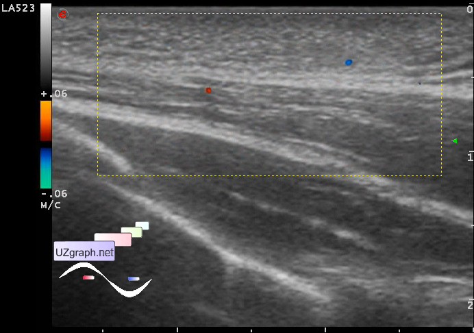
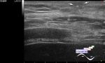
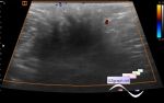
    A child 6 months old after cryoablation of skin hemangioma in the left side of the chest.

    Visually in this area mostly white-gray circular spot with a bluish area which a little above the level of intact skin (a condition after cryoablation, scar).

    At the US in the area of the scar visualized moderately inhomogeneous mostly hyperechoic area with an unclear contour up to 2cm wide.

    By the words of the accompanying, cryosurgery was performed gradually, by pieces, probably this explains the heterogeneity, ie partial destruction of hemangioma.

    external link

:: attachments(4) ::

:: file 1 ::

:: file 2 ::

:: file 3 ::

:: file 4 ::


 Random 
Ultrasound cases - DCM vs NCM
    

Ultrasound cases - Testicular microlith
    

Ultrasound cases - Not a Rubik's Cubes
    

Ultrasound cases - Linea alba hernia vs lipoma part deux
    

Ultrasound cases - Testicle in the anterior abdominal wall
    

 Dictionary 
MSD

RVOT

EFW

PRF

TAMEAN

SF

IVSd

Color M-mode

Freeze

LVESD


 Calculators 
Pressure gradient

Estimated fetal weight by Ultrasound

Thyroid volume

Body surface area

Teicholz systolic cardiac function


 Similar topics 
Ultrasound cases - Penetrating injury of the femur
    

Ultrasound cases - Cephalohematoma
    

Ultrasound cases - Urachus fistula
    

Ultrasound cases - Cranial mass
    

Ultrasound cases - Pilonidal coccyx cyst
    

Ultrasound cases - Palm fistula, in search of pencil lead
    

Ultrasound cases - Postoperative abdominal wall fistula
    

Ultrasound cases - Nucks cyst
    

Ultrasound cases - Paraproctitis
    

Ultrasound cases - Submandibular area lesion