Log in    RU  


Main page :: Forum :: Ultrasound cases - Sonograms, cine-loops etc. ::

Large Ventricular Septal Defect (VSD)


Tags: Cardiac sonography(Echocardiography), GE Logiq Book XP, Clinical report, Pediatric

   Posts
 
  Large Ventricular Septal Defect (VS...
#1

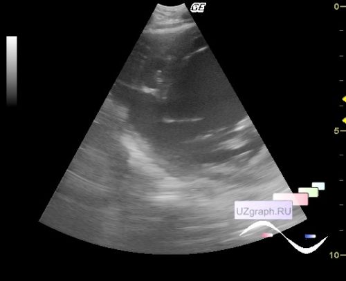
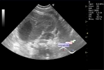
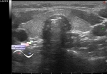
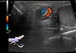
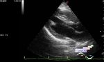
    A 2-year-old child in the ICU of the Children's City Clinical Hospital with congenital heart disease (CHD, large VSD, mainly posterior septal segments, which is clearly visible on views along the short axis) and with deformity of the chest (due to which not all echocardiography views can be obtained).


 Random 
Ultrasound cases - Lesion in the projection of the adrenal gland
    

Ultrasound cases - 2 plus 2
    

Ultrasound cases - Rested
    

Ultrasound cases - Appendicular infiltrate
    

Ultrasound cases - Clinically suspected appendicitis
    

 Dictionary 
LVESV

Qp-Qs

PG

SV

Color M-mode

ESV

RAP

FVI

Freeze

LN


 Calculators 
Thyroid volume

Pressure gradient

Estimated fetal weight by Ultrasound

Teicholz systolic cardiac function

TI-RADS


 Similar topics 
Ultrasound cases - Bicuspid aortic valve 2
    

Ultrasound cases - Mitral valve cyst
    

Ultrasound cases - Electrode in the right heart after installing of heart pacemaker
    

Ultrasound cases - Chylopericardium, lung atelectasis, cardiomegaly, ascites
    

Ultrasound cases - Asymmetric LVH, aortic regurgitation
    

Ultrasound cases - Coronary fistula from LCA?
    

Ultrasound cases - Floating IAS aneurysm
    

Ultrasound cases - Aortic Stenosis
    

Ultrasound cases - Floating Accessory Pulmonary Valve Tissue
    

Ultrasound cases - Hyperechoic linear structure in the left atrium