Log in    RU  


Main page :: Forum :: Ultrasound cases - Sonograms, cine-loops etc. ::

Flotation in the lateral ventricle


Tags: Brain sonography(Neurosonography), GE Logiq E, Clinical report, Pediatric

   Posts
 
  Flotation in the lateral ventricle
#1

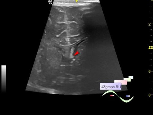
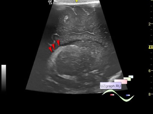
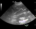
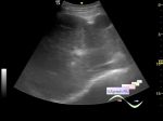
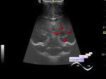
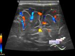
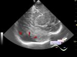
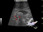
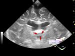
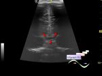
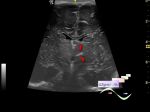
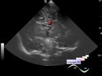
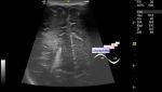
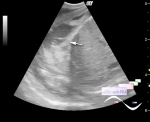
    Infant 1 month old came to the public clinic for a scheduled echo screening.

    On the NSG in the projection of the right lateral ventricle, an inclusion is visualized as a complex cyst up to 4x5x6mm (subependymal cyst). In the left ventricle, an echogenic floating structure is visualized visually fixed to the choroid plexus (differential diagnosis: intraventricular hemorrhage / thrombus, etc.)


    external link

  04:44 17-01-2023
 
#2

    More about flotation -

    external link


 Random 
Scientific - Playlist Intestinal ultrasound

Ultrasound cases - DCM vs NCM
    

Ultrasound cases - Ileotyphlitis and/or ileocecal intussusception
    

Ultrasound cases - Postoperative ureteritis
    

Ultrasound cases - Liver cirrhosis and thickened omentum
    

 Dictionary 
Ved

LVPWd

HR

EDV

Gain

Doppler angle

Aliasing doppler artifact

FL

IVSd

CRL


 Calculators 
Estimated fetal weight by Ultrasound

Pulmonary artery pressure

Qp:Qs

Gestational age by Ultrasound

Thyroid volume


 Similar topics 
Ultrasound cases - Cyst in the projection near the posterior horn of the lateral ventricle
    

Ultrasound cases - Subdural collection
    

Ultrasound cases - Anechogenic inclusion in the posterior cranial fossa
    

Ultrasound cases - Cyst of the base of the brain
    

Ultrasound cases - Interthalamic adhesion aberration
    

Ultrasound cases - Suprasellar lession
    

Ultrasound cases - Hyperechoic lesion in the thalamus
    

Ultrasound cases - Chord of the brain lateral ventricle
    

Ultrasound cases - Increased periventricular echogenicity
    

Ultrasound cases - One and a half kilograms of happiness