Log in    RU  


Main page :: Forum :: Ultrasound cases - Sonograms, cine-loops etc. ::

Increased periventricular echogenicity


Tags: Brain sonography(Neurosonography), GE Logiq E, Clinical report, Pediatric

   Posts
 
  Increased periventricular echogenic...
#1

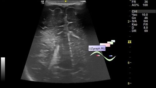
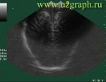
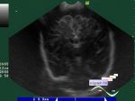
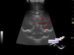
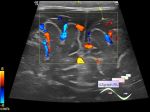
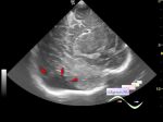
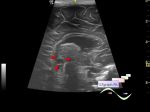
    Child 1 month age in the public clinic was sent for a planned neurosonography.

    At ultrasound visualized an increase of periventricular echogenicity on both sides (differential diagnosis: immaturity, periventricular leukomalacia, etc.).

    A follow-up ultrasound is recommended after 1 month.


 Random 
Ultrasound cases - Clinically suspected appendicitis
    

Admin room - International day of radiology

Ultrasound cases - Clinic is always right
    

Ultrasound cases - Infant normal adrenal
    

Ultrasound cases - Vertebral artery hypoplasia in adolescents
    

 Dictionary 
Anatomical M-mode

Freeze

EF

CFM

GS

DPD

BPD

HR

LVESV

LN


 Calculators 
Pulmonary artery pressure

Pressure gradient

Volume (ellipse formula)

Thyroid volume

Resistive index


 Similar topics 
Ultrasound cases - Echogenic brain foci
    

Ultrasound cases - Subdural hemorrhage 2
    

Ultrasound cases - Willis circle at ultrasound
    

Ultrasound cases - Periventricular hyperechogenicity
    

Ultrasound cases - Combined (internal and external) hydrocephalus
    

Ultrasound cases - Subdural hemorrhage
    

Ultrasound cases - Cyst in the projection near the posterior horn of the lateral ventricle
    

Ultrasound cases - Subdural collection
    

Ultrasound cases - Anechogenic inclusion in the posterior cranial fossa
    

Ultrasound cases - Cyst of the base of the brain