Log in    RU  


Main page :: Forum :: Ultrasound cases - Sonograms, cine-loops etc. ::

Acne on the back


Tags: Soft tissues sonography, Images, Video, Clinical report, Esaote MyLab 70, Adult

   Posts
 
  Acne on the back
#1

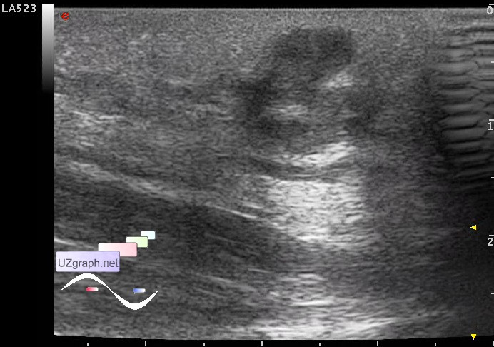
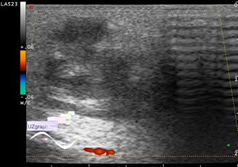
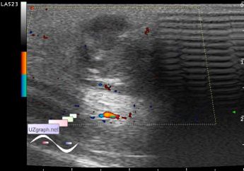
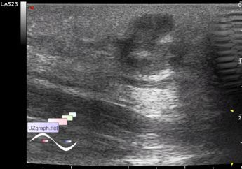
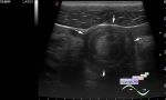
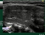
    The patient 50 years-old with complaints of lesion on the back. Visually, prominent above the skin, up to 2.5 cm in diameter, red color, in history there was a such lesion which was underwent surgery and now represented as a rough scar (by the words of patient the surgeon did not sew).

    At ultrasound hypoechoic lesion of irregular shape up to 10x5mm, vertical orientation, with no blood flow at DPD, surrounded by hyperechoic tissue with increased blood flow (dif.diagnosis: atheroma/sebaceous cyst, etc.).

    external link

:: attachments(6) ::

:: file 1 ::

:: file 2 ::

:: file 3 ::

:: file 4 ::

:: file 5 ::

:: file 6 ::


 Random 
Ultrasound cases - Area with hyperechoic dot inclusions
    

Ultrasound cases - Combined (internal and external) hydrocephalus
    

Ultrasound cases - Bifurcated rib (Lushka's fork)
    

Ultrasound cases - Small muscular interventricular septum shunts
    

Ultrasound cases - Bilateral popliteal cysts with legs
    

 Dictionary 
PADP

LVESD

SV

Doppler angle

ESV

LVPWd

Ved

HC

TVUS

TAV


 Calculators 
Type of an infant hip joint

Pulmonary artery pressure

Estimated fetal weight by Ultrasound

Gestational age by Ultrasound

Body surface area


 Similar topics 
Ultrasound cases - Penetrating injury of the femur
    

Ultrasound cases - Cephalohematoma
    

Ultrasound cases - Urachus fistula
    

Ultrasound cases - Cranial mass
    

Ultrasound cases - Pilonidal coccyx cyst
    

Ultrasound cases - Palm fistula, in search of pencil lead
    

Ultrasound cases - Postoperative abdominal wall fistula
    

Ultrasound cases - Nucks cyst
    

Ultrasound cases - Paraproctitis
    

Ultrasound cases - Submandibular area lesion