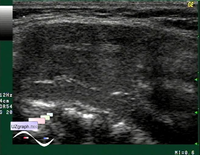
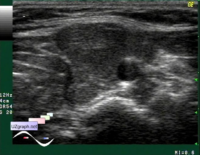
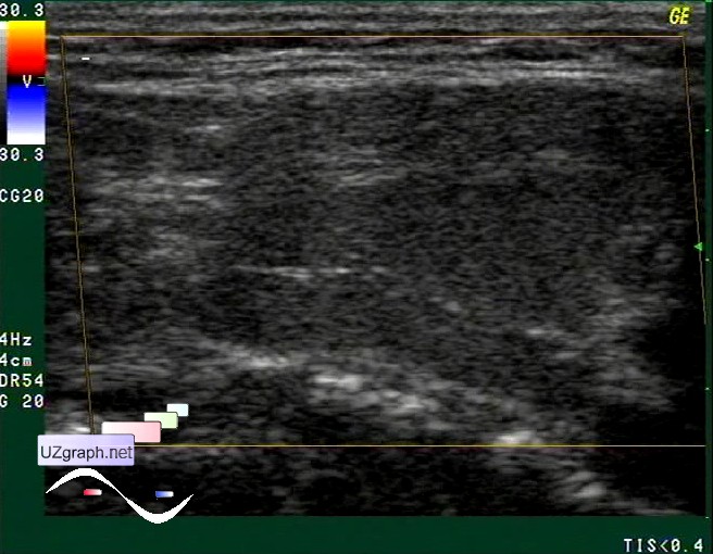
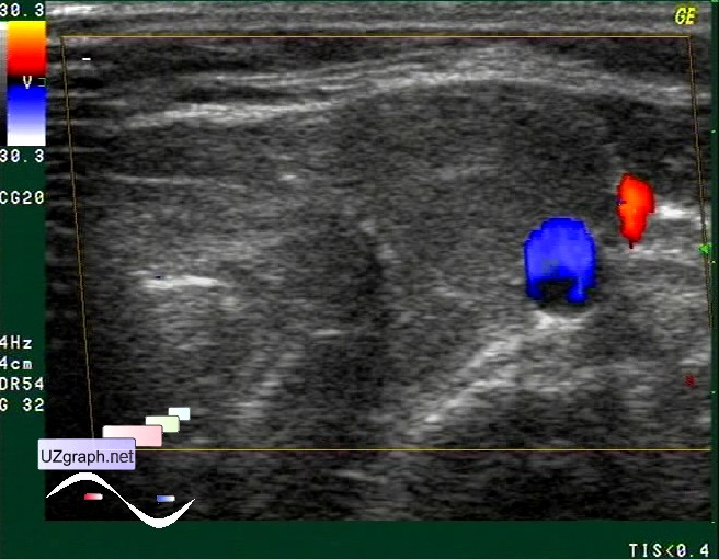
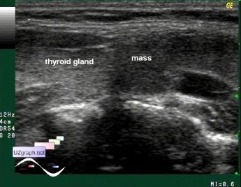
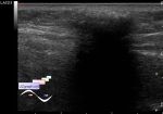
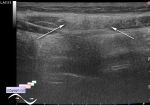
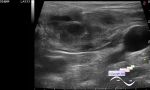
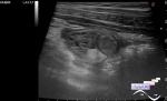
Log in    RU  


Main page :: Forum :: Ultrasound cases - Sonograms, cine-loops etc. ::

Neck lymphangioma


Tags: Soft tissues sonography, Images, Video, Clinical report, GE Logiq 400 MD, Pediatric

   Posts
 
  Neck lymphangioma
#1

    Teenager with visible side neck mass.

    At ultrasound at first look mass simulate solid structure but upon a closer view it' s a complex cyst with sludge positioned close to thyroid gland and neck vessels.

    Histologically verified as lymphangioma.

    external link

:: attachments(6) ::

:: file 1 ::

:: file 2 ::

:: file 3 ::

:: file 4 ::

:: file 5 ::

:: file 6 ::


 Random 
Ultrasound cases - Testicular torsion after surgery and thyroid
    

Ultrasound cases - Ascariasis of small intestine
    

Ultrasound cases - "Tricky" IVSD
    

Ultrasound cases - MV fistula?
    

Ultrasound cases - Dolichosigma
    

 Dictionary 
PSV

EF

LN

FL

HR

TAV

GA

Freeze

PA

Color M-mode


 Calculators 
Qp:Qs

Volume (ellipse formula)

Teicholz systolic cardiac function

Estimated fetal weight by Ultrasound

Thyroid volume


 Similar topics 
Ultrasound cases - Skin shell
    

Ultrasound cases - Skin lesion in the projection of the surgical suture
    

Ultrasound cases - Purulent / Suppurative lymphadenitis
    

Ultrasound cases - Hematoma of the gluteal region
    

Ultrasound cases - Thickening and heterogeneity of the intercostal space soft tissues
    

Ultrasound cases - Chest wall lesion
    

Ultrasound cases - Hockey Elbow
    

Ultrasound cases - Combination of left shoulder lesions
    

Ultrasound cases - Infiltrate/ phlegmon of the wrist
    

Ultrasound cases - Subcutaneous lesion above the sternum