DCM vs NCM
Tags: Cardiac sonography(Echocardiography), Esaote MyLab 70, Images, Video, Clinical report, Pediatric
| Posts | |||
| DCM vs NCM | #1 |
| |||||
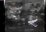
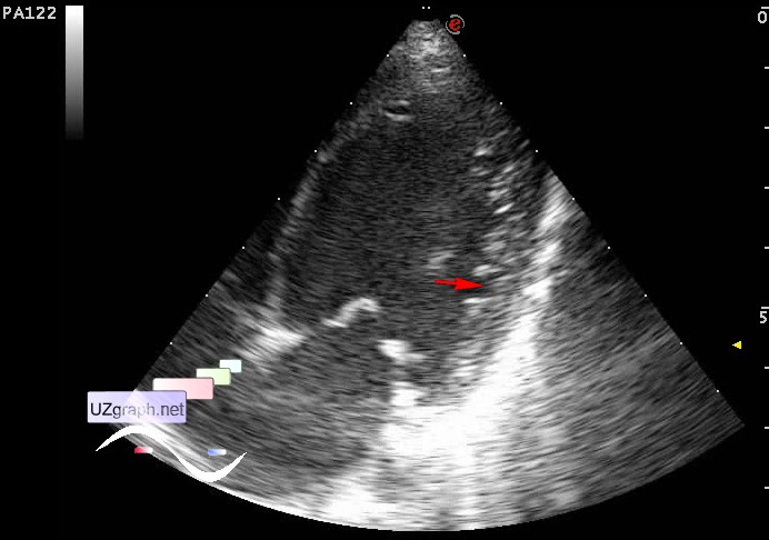
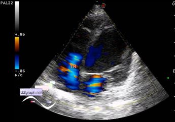
:: file 1 ::
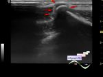
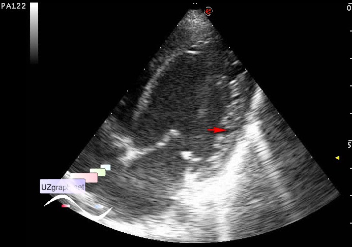
 :: file 2 ::
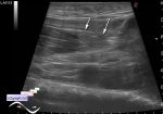
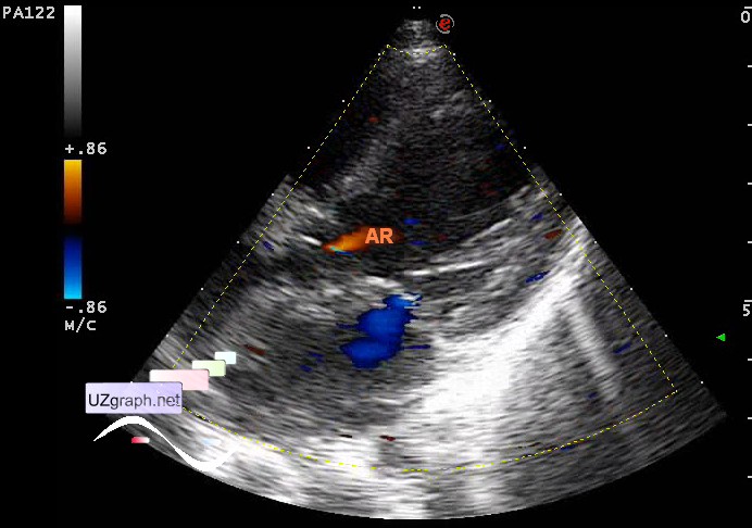
 :: file 3 ::
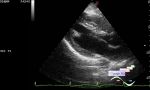
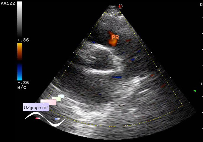
 :: file 4 ::
 :: file 5 ::
 :: file 6 ::
 :: file 7 :: | |||||


| 14:13 25-10-2015 | #2 |
| |||||