Log in    RU  


Main page :: Forum :: Ultrasound cases - Sonograms, cine-loops etc. ::

Scapula exostosis


Tags: Musculoskeletal sonography, Soft tissues sonography, GE Logiq E, Clinical report, Pediatric

   Posts
 
  Scapula exostosis
#1

    A 7-year-old child in the public clinic with complaints of a mass in the projection of the scapula was referred for an ultrasound of soft tissues, an X-ray was previously performed where, according to the words of the accompanying child, nothing was found.

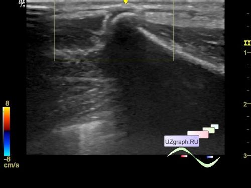
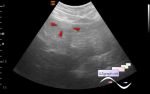
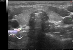
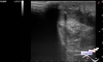
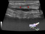
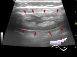
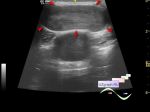
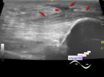
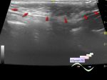
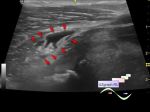
    In the projection of the mass visible to the naked eye in the projection of the right scapula, moving along with the scapula, at ultrasound visualizes a hyperechoic lesion with an acoustic shadow, intimately located along the edge of the scapula, without blood flow at CFM, up to 7 mm in size (differential diagnosis: exostosis, etc.).


 Random 
Ultrasound cases - Lymphadenopathy of the cervical lymph nodes
    

Ultrasound cases - Hirschsprung disease on ultrasound
    

Ultrasound cases - Tetralogy of Fallot
    

Ultrasound cases - Parietal bladder lesion
    

Ultrasound cases - Situs inversus
    

 Dictionary 
HR

CO

SV

DPD

PASP

EFW

Gain

MC

RAP

PD


 Calculators 
Estimated fetal weight by Ultrasound

Thyroid volume

Volume (ellipse formula)

Gestational age by Ultrasound

Myocardial mass


 Similar topics 
Ultrasound cases - Penetrating injury of the femur
    

Ultrasound cases - Cephalohematoma
    

Ultrasound cases - Cranial mass
    

Ultrasound cases - Recurrent shin trauma
    

Ultrasound cases - Knee joint synovitis
    

Ultrasound cases - Thickening and heterogeneity of the intercostal space soft tissues
    

Ultrasound cases - Chest wall lesion
    

Ultrasound cases - Hockey Elbow
    

Ultrasound cases - Combination of left shoulder lesions
    

Ultrasound cases - Baker's cyst and intramuscular hematoma