Log in    RU  


Main page :: Forum :: Ultrasound cases - Sonograms, cine-loops etc. ::

Cysts in the pelvis


Tags: Female pelvis sonography, Medison Sonoace R7, Images, Video, Clinical report, Pediatric

   Posts
 
  Cysts in the pelvis
#1

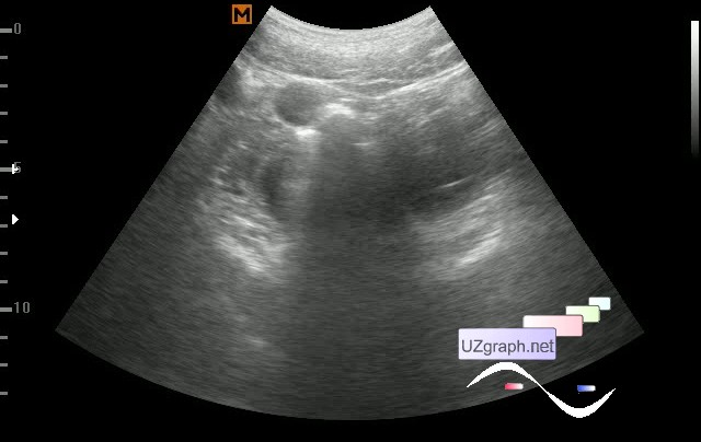
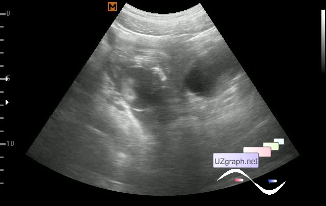
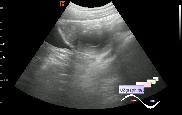
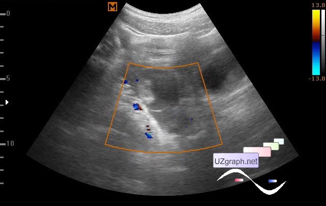
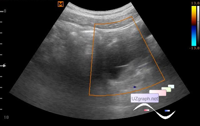
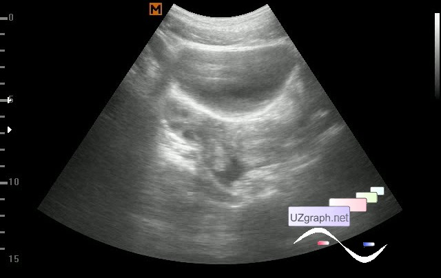
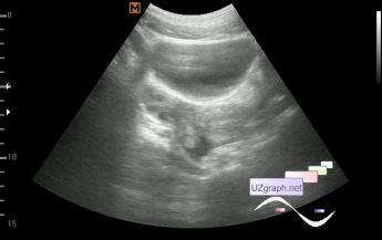
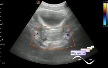
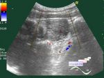
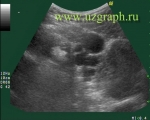
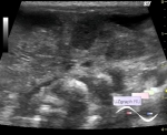
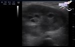
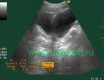
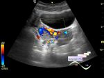
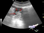
    Child 16 years old, several months have been observed with ovarian cysts.

    On ultrasound in both ovaries there are cysts. Both ovary are enlarged, right in size up to 68x38mm, with cysts, in one of them a parietal formation of a solid type up to 3 cm (teratoma? hemorrhagic cyst?), Left to 52x37mm, is represented by a cyst with septa (hemorrhagic cyst?)

    Attachments 1-6.

    The child was directed to the child hospital and surgery was performed. Laparoscopic operation was performed: cystectomy on the right, removal of parovarial cyst on the right, removal of paratubar cyst on the right.

    As far as I understood from the words of the gynecologist and the patient, on the histology of the cyst of the right ovary is described as a cyst of the yellow body, which caused doubts both in me and in the gynecologist. The cyst persisted for a long time and did not react to hormonal treatment.

    In the postoperative ultrasound at hospital was indicated the presence of free fluid in the Douglas space up to 55ml.

    After about a month after surgery at follow-up ultrasound in clinic, cysts in the ovaries are not determined, in the Douglas space to 21 ml of free fluid, more to the right. Also between the uterus and the right ovary is visualized an echogenic lesion up to 2 cm (corpus alienum (hemostatic sponge)?).

    Attachments 7-9.

    external link

:: attachments(9) ::

:: file 1 ::

:: file 2 ::

:: file 3 ::

:: file 4 ::

:: file 5 ::

:: file 6 ::

:: file 7 ::

:: file 8 ::

:: file 9 ::


 Random 
Ultrasound cases - Situs inversus
    

Technical - Micro-ultrasound

Ultrasound cases - Thickening of the sternocleidomastoid
muscle


Ultrasound cases - Choroid plexus cyst
    

Ultrasound cases - Megaureter, ureteral valve-stone
    

 Dictionary 
Doppler angle

PADP

LVPWd

LVEDD

M-mode

LMP

PG

SV

LVOT

TVUS


 Calculators 
Type of an infant hip joint

Pulmonary artery pressure

Qp:Qs

Myocardial mass

Body surface area


 Similar topics 
Ultrasound cases - Nucks cyst
    

Ultrasound cases - Hydrosalpinx (saktosalpinx)
    

Ultrasound cases - Portal vein air in the newborn
    

Ultrasound cases - Ovarian teratoma
    

Ultrasound cases - Inguinal hernia with an ovary
    

Ultrasound cases - Giant cyst of the right lateral canal
    

Ultrasound cases - Right ovary changes
    

Ultrasound cases - Ovarian apoplexy 3
    

Ultrasound cases - Hemorrhagic ovarian cyst
    

Ultrasound cases - Patchy uterus