Log in    RU  


Main page :: Forum :: Ultrasound cases - Sonograms, cine-loops etc. ::

Liver hemangioma plus


Tags: Abdomen sonography, GE Vivid 3, Images, Video, Clinical report, Adult

   Posts
 
  Liver hemangioma plus
#1

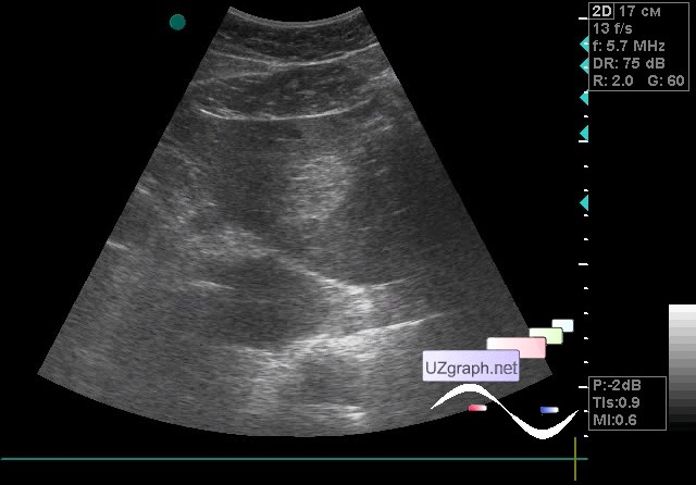
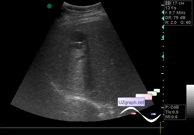
    A 55-year-old patient came to the control of an abdomen ultrasound scan, in the previous ultrasound scan report in the left lobe of liver noticed the hemangioma.

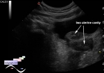
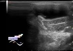
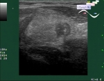
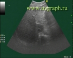
    On the current ultrasound, visualization of the liver, especially of the right lobe, is difficult / limited; performed at a deep breath through the intercostal spaces by fragments: in the left hepatic lobe the hyperechoic lesion up to 4x2.5 cm is visualized; and a group of cysts or a multi-chamber cyst in the 5-6th segment of the liver also visualized(echinococcosis? other?)

    external link

:: attachments(3) ::

:: file 1 ::

:: file 2 ::

:: file 3 ::


 Random 
Ultrasound cases - Bladder mass
    

Ultrasound cases - Liver pseudotumor
    

Ultrasound cases - Clinically suspected appendicitis
    

Ultrasound cases - Megacolon
    

Ultrasound cases - Spinal surgery complication
    

 Ad 


 Dictionary 
CFM

TV

FVI

HR

Qp-Qs

EF

EDV

HC

LVPWd

AC


 Calculators 
Qp:Qs

Myocardial mass

Body surface area

Type of an infant hip joint

Estimated fetal weight by Ultrasound


 Similar topics 
Ultrasound cases - Hirschsprung disease on ultrasound
    

Ultrasound cases - Intestinal Infiltrate
    

Ultrasound cases - Abdominal cryptorchidism
    

Ultrasound cases - Diffuse heterogeneous liver
    

Ultrasound cases - Postoperative abdominal wall fistula
    

Ultrasound cases - Stump appendicitis
    

Ultrasound cases - Liver scar after resection
    

Ultrasound cases - Subdiaphragmatic movable fluid collection (UFO)
    

Ultrasound cases - Lesion of the spleen or the case when it is easy to not notice
    

Ultrasound cases - I’m normal but others ...