Log in    RU  


Main page :: Forum :: Ultrasound cases - Sonograms, cine-loops etc. ::

Left inferior vena cava


Tags: Vascular sonography, GE Logiq 400 MD, Images, Video, Clinical report, Pediatric

   Posts
 
  Left inferior vena cava
#1

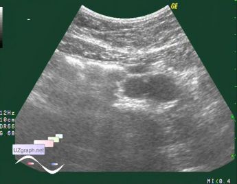
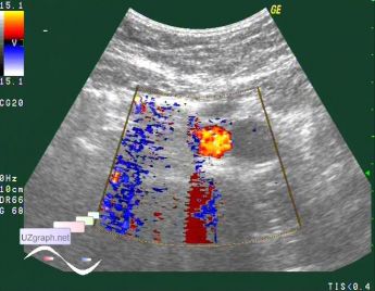
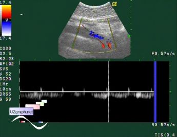
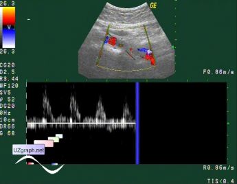
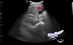
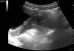
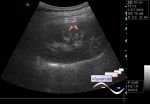
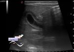
    Routine abdominal ultrasound of teenager with abdominal pain.

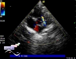
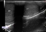
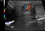
    In the mid-abdomen visualized atypical two vessels view (files 1,2) - to the left of the aorta closely to it second vessel with venous doppler spectrum(file 3), reverse to aorta spectrum(file 4).

    external link

:: attachments(5) ::

:: file 1 ::

:: file 2 ::

:: file 3 ::

:: file 4 ::

:: file 5 ::


 Random 
Ultrasound cases - Inguinal hernia with an ovary
    

Ultrasound cases - Strange RVOT
    

Ultrasound cases - Choledochal cyst
    

Admin room - Ultrasound Flash revolution
    

Ultrasound cases - Small muscular interventricular septum shunts
    

 Dictionary 
Qp-Qs

EF

IVSd

HC

MSD

ESV

PG

ESD

FVI

Ved


 Calculators 
Thyroid volume

Body surface area

Pressure gradient

Teicholz systolic cardiac function

Type of an infant hip joint


 Similar topics 
Ultrasound cases - Subcutaneous abnormal body vessel
    

Ultrasound cases - Complication of a blood test from a vein
    

Ultrasound cases - Anomaly (doubling) of the popliteal vein
    

Ultrasound cases - Varicose disease
    

Ultrasound cases - Vertebral artery hypoplasia in adolescents
    

Ultrasound cases - RLQ phlebectasia
    

Ultrasound cases - Anomaly of caudal aorta
    

Ultrasound cases - Varicose and Baker's cyst
    

Ultrasound cases - Varicose veins of LE
    

Ultrasound cases - Cubital vein thrombosis