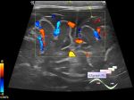
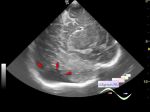
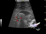
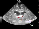
Log in    RU  


Main page :: Forum :: Ultrasound cases - Sonograms, cine-loops etc. ::

Double - IVH


Tags: Brain sonography(Neurosonography), Cardiac sonography(Echocardiography), Abdomen sonography, Medison Sonoace R7, Images, Video, Clinical report, Pediatric

   Posts
 
  Double - IVH
#1

    Children 2 months old, from twin pregnancy. Perinatally suffered an IVH (intraventricular hemorrhage), pneumonia, were in intensive care unit.

    At one in connection with ventriculomegaly occurred after IVH, has several punctures of the LV (lateral ventricles) and then a VPS (ventriculoperitoneal shunt) was performed, a CT scan revealed a periventricular/connatal cysts (PCs), on NSG (neurosonography) before CT, no cysts were detected (I also did not immediately notice them). At this moment, the transverse size of the LV is up to 15 mm each, in the lower parts of the occipital horns small thrombi are visible.

    At the second the PCs was identified at the NSG.

    Both of them currently have PDA (patent ductus arteriosus).

    The second also has the SEC (subependimal cyst) and a bile clot of the gallbladder.

    external link

:: attachments(16) ::

:: file 1 ::

:: file 2 ::

:: file 3 ::

:: file 4 ::

:: file 5 ::

:: file 6 ::

:: file 7 ::

:: file 8 ::

:: file 9 ::

:: file 10 ::

:: file 11 ::

:: file 12 ::

:: file 13 ::

:: file 14 ::

:: file 15 ::

:: file 16 ::

  00:59 11-10-2021
  Doble - HIV, CAP, etc.
#2

    Niños de 2 meses, desde gestación gemelar. Sufrió perinatalmente una hemorragia intraventricular (HIV), neumonía, se encontraba en la unidad de cuidados intensivos.

    Uno tiene cortocircuito ventriculoperitoneal debido a ventriculomegalia severa, en las partes inferiores de los cuernos occipitales son visibles pequeños trombos.

    Ambos tienen CAP (conducto arterioso persistente) y quistes periventriculares / connatales.

    Otro tiene un quiste subependimal y un coágulo de bilis en la vesícula biliar.

    external link


 Random 
Ultrasound cases - Partial tears of the lateral meniscus
    

Ultrasound cases - Position anomalies of the spleen and left kidney
    

Ultrasound cases - Pyelitis, cystitis
    

Ultrasound cases - All the small things...
    

On-call room - And what is your gel, sterile?

 Dictionary 
B-mode

MSD

LVESD

LN

CO

LVEDV

Gain

BPD

TVUS

IVSd


 Calculators 
Pulmonary artery pressure

Resistive index

Myocardial mass

Gestational age by Ultrasound

Volume (ellipse formula)


 Similar topics 
Ultrasound cases - Cyst in the projection near the posterior horn of the lateral ventricle
    

Ultrasound cases - Subdural collection
    

Ultrasound cases - Anechogenic inclusion in the posterior cranial fossa
    

Ultrasound cases - Cyst of the base of the brain
    

Ultrasound cases - Interthalamic adhesion aberration
    

Ultrasound cases - Suprasellar lession
    

Ultrasound cases - Flotation in the lateral ventricle
    

Ultrasound cases - Hyperechoic lesion in the thalamus
    

Ultrasound cases - Chord of the brain lateral ventricle
    

Ultrasound cases - Increased periventricular echogenicity