Log in    RU  


Forum :: Ultrasound cases - Sonograms, cine-loops etc. ::

Necrotizing Enterocolitis


Replies: 0   Rating: none

Tags: Abdomen sonography, Gastrointestinal sonography, GE Logiq E, Clinical report, Pediatric

Moderator :
   User: admin
   Registered: 23-09-2013
   Posts: 892
   Name: Kolesnichenko Yuri
   Profession: MD, Sonologist
   City: Moscow, Russia
   New reply  
   Posts
  07:44 20-01-2023
  Necrotizing Enterocolitis
#1
   User: admin
   Registered: 23-09-2013
   Posts: 892
   Name: Kolesnichenko Yuri
   Profession: MD, Sonologist
   City: Moscow, Russia

    A 2-year-old child with complaints of abdominal pain was urgently sent for an ultrasound scan in a public clinic.

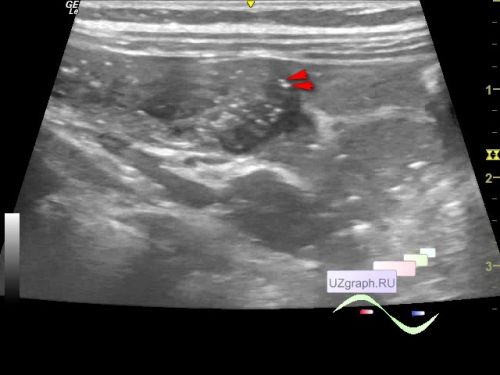
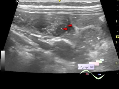
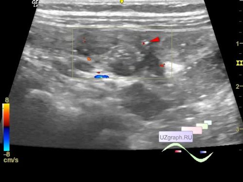
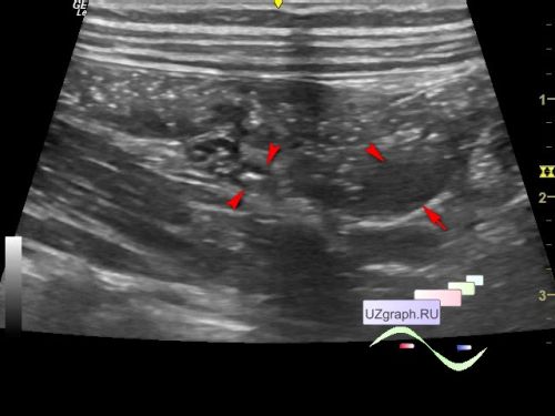
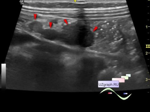
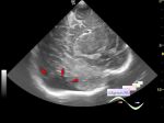
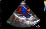
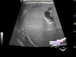
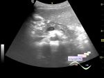
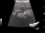
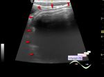
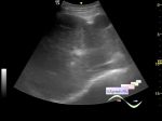
    On ultrasound in the right iliac region, the caecum with a wall thickened up to 5 mm and hyperechoic microinclusions in the wall is visualized (differential diagnosis: typhlitis with pneumatosis of the intestinal wall - Pneumatosis intestinalis — gas in the bowel wall / necrotizing enterocolitis, etc.). More than 5 lymph nodes up to 12 mm are visualized in the mesogastrium (differential diagnosis: mesadenitis, etc.). In the right iliac region, a tubular structure up to 3 mm in thickness is visualized (differential diagnosis: normal appendix, etc.)

    An urgent consultation with an infectious disease specialist is recommended.


Video from Youtube

WYSIWYG for Geek
   Qu.  
   Edit  
   Del.  
   Access  
 
   New reply  


 Ad 

 Random 
Ultrasound cases - Osgood-Schlatter disease, tendinitis
    

Ultrasound cases - Anechogenic inclusion in the posterior cranial fossa
    

Ultrasound cases - Postoperative abdominal infiltrate
    

Ultrasound cases - Linea alba hernia vs lipoma part deux
    

Ultrasound cases - Additional flow to LVOT
    

 Ad 


 Dictionary 
LN

Anatomical M-mode

Aliasing doppler artifact

PD

M-mode

Freeze

CW

Color M-mode

Doppler angle

TVUS


 Similar topics 
Ultrasound cases - Cholelithiasis and gallbladder cholesterosis
    

Ultrasound cases - Portal vein spontaneous enhancement (mononucleosis)
    

Ultrasound cases - Hypoechoic spleen lesion
    

Ultrasound cases - Acute Appendicitis, periappendicular infiltrate
    

Ultrasound cases - Cavernous Transformation of the Portal Vein
    

Ultrasound cases - Diffuse-focal changes in the liver, thick left kidney
    

Ultrasound cases - Lesion in the projection of the pancreas body
    

Ultrasound cases - Double barrel shotgun sign
    

Ultrasound cases - Megacolon
    

Ultrasound cases - Liver cirrhosis and thickened omentum


  © Uzgraph.net 2013-22
  © Uzgraph.ru 2008-23