Log in    RU  


Main page :: Forum :: Ultrasound cases - Sonograms, cine-loops etc. ::

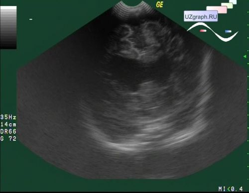
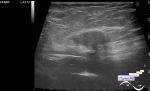
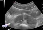
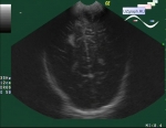
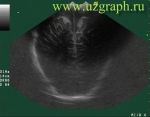
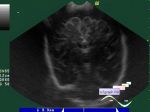
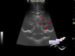
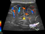
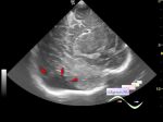
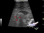
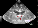
Combined (internal and external) hydrocephalus


Tags: Brain sonography(Neurosonography), GE Logiq 400 MD, Clinical report, Pediatric

   Posts
 
  Combined (internal and external) hy...
#1

    A baby in a public clinic at a neurosonography.

    At Ultrasound of the brain visualizes the dilatation of the brain lateral ventricles and subconvexital space.


 Random 
Ultrasound cases - Olecranon bursitis
    

Ultrasound cases - Electrode in the right heart after installing of heart pacemaker
    

Ultrasound cases - Such different ovaries
    

Ultrasound cases - Cryptorchidism, testicular hypoplasia
    

Ultrasound cases - Clinically suspected appendicitis
    

 Dictionary 
TVUS

LVOT

TI-RADS

RAP

AC

BSA

EDV

FNA

ESD

EFW


 Calculators 
TI-RADS

Pulmonary artery pressure

Thyroid volume

Teicholz systolic cardiac function

Type of an infant hip joint


 Similar topics 
Ultrasound cases - Echogenic brain foci
    

Ultrasound cases - Subdural hemorrhage 2
    

Ultrasound cases - Willis circle at ultrasound
    

Ultrasound cases - Periventricular hyperechogenicity
    

Ultrasound cases - Subdural hemorrhage
    

Ultrasound cases - Cyst in the projection near the posterior horn of the lateral ventricle
    

Ultrasound cases - Subdural collection
    

Ultrasound cases - Anechogenic inclusion in the posterior cranial fossa
    

Ultrasound cases - Cyst of the base of the brain
    

Ultrasound cases - Interthalamic adhesion aberration