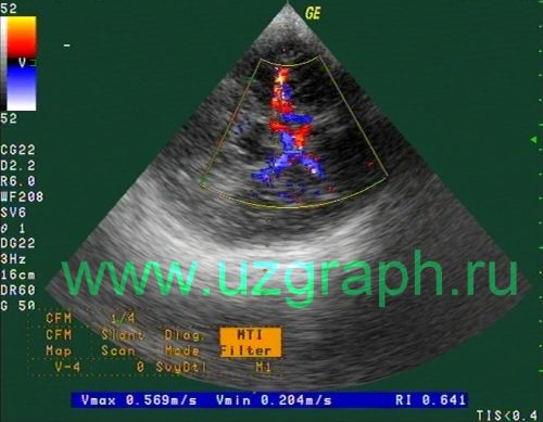
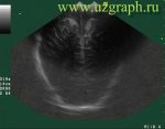
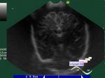
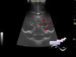
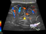
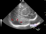
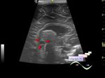
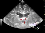
Log in    RU  


Main page :: Forum :: Ultrasound cases - Sonograms, cine-loops etc. ::

Willis circle at ultrasound


Tags: Brain sonography(Neurosonography), GE Logiq 400 MD, Clinical report, Pediatric

   Posts
 
  Willis circle at ultrasound
#1

    An infant in a public clinic undergoing NSG with TCD.


 Random 
Ultrasound cases - Old prepatellar bursitis
    

Ultrasound cases - Linea alba hernia vs lipoma part deux
    

Ultrasound cases - Good vs Evil - thyroid 4
    

Ultrasound cases - Inguinal hernia, hydrocele
    

Ultrasound cases - PO suture
    

 Dictionary 
CFM

B-mode

PD

MC

PASP

IVSd

TV

EFW

LVEDV

BPD


 Calculators 
TI-RADS

Volume (ellipse formula)

Resistive index

Gestational age by Ultrasound

Type of an infant hip joint


 Similar topics 
Ultrasound cases - Echogenic brain foci
    

Ultrasound cases - Subdural hemorrhage 2
    

Ultrasound cases - Periventricular hyperechogenicity
    

Ultrasound cases - Combined (internal and external) hydrocephalus
    

Ultrasound cases - Subdural hemorrhage
    

Ultrasound cases - Cyst in the projection near the posterior horn of the lateral ventricle
    

Ultrasound cases - Subdural collection
    

Ultrasound cases - Anechogenic inclusion in the posterior cranial fossa
    

Ultrasound cases - Cyst of the base of the brain
    

Ultrasound cases - Interthalamic adhesion aberration