Log in    RU  


Main page :: Forum :: Ultrasound cases - Sonograms, cine-loops etc. ::

Strange RVOT


Tags: Cardiac sonography(Echocardiography), GE Logiq P6, Images, Video, Clinical report, Pediatric

   Posts
 
  Strange RVOT
#1

    Teenager examined because of complaints about attacks accompanied by weakness and blackout in the eyes.

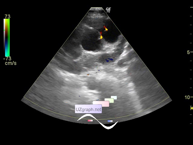
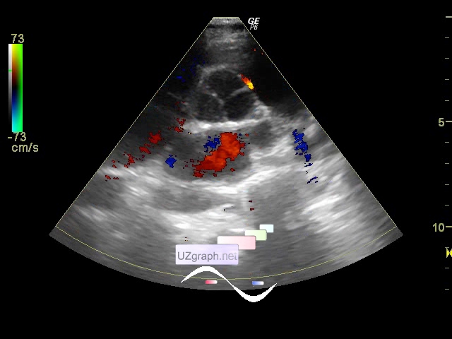
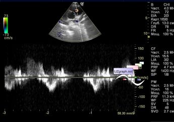
    At first glance, the heart is normal, drew attention to the uneven heart rate, small AR, but then noticed an interesting picture in a projection of RVOT - 2 reversal jets, one of which projected classically in the field of clamping leaflets PV, and the second in the projection of the mouth of the left coronary artery (coronary fistula?)

:: attachments(6) ::

:: file 1 ::

:: file 2 ::

:: file 3 ::

:: file 4 ::

:: file 5 ::

:: file 6 ::

  16:57 23-11-2014
 
#2

    Similar picture:

    "Coronary artery-pulmonary artery fistula: case report":

    external link

  10:08 17-06-2015
 
#3

    Similar picture, ALCAPA case:

    external link


 Random 
Ultrasound cases - Cystic dysplasia, hematuria
    

Ultrasound cases - Tetralogy of Fallot
    

Ultrasound cases - nabothian cyst
    

Ultrasound cases - Diffuse-focal changes in the liver, thick left kidney
    

Ultrasound cases - Torticollis
    

 Dictionary 
CO

LVEDV

TVUS

AC

LVEDD

IVC

PA

CRL

HR

LN


 Calculators 
Body surface area

Qp:Qs

Teicholz systolic cardiac function

TI-RADS

Volume (ellipse formula)


 Similar topics 
Ultrasound cases - Bicuspid aortic valve 2
    

Ultrasound cases - Mitral valve cyst
    

Ultrasound cases - Electrode in the right heart after installing of heart pacemaker
    

Ultrasound cases - Chylopericardium, lung atelectasis, cardiomegaly, ascites
    

Ultrasound cases - Asymmetric LVH, aortic regurgitation
    

Ultrasound cases - Coronary fistula from LCA?
    

Ultrasound cases - Floating IAS aneurysm
    

Ultrasound cases - Aortic Stenosis
    

Ultrasound cases - Floating Accessory Pulmonary Valve Tissue
    

Ultrasound cases - Hyperechoic linear structure in the left atrium