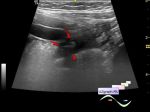
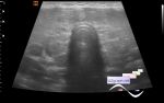
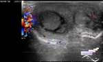
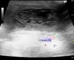
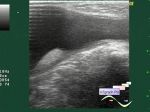
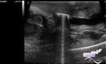
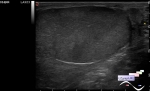
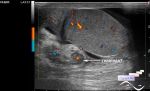
Log in    RU  


Main page :: Forum :: Ultrasound cases - Sonograms, cine-loops etc. ::

Cryptorchidism


Tags: Scrotum sonography, Images, Video, Clinical report, Esaote MyLab 70, Pediatric

   Posts
 
  Cryptorchidism
#1

    Child after surgeon' s examination aimed at ultrasound with diagnosis of hypoplasia of both testicles.

    Visually, scrotum is empty.

    On US testicles in the proximal third of the inguinal canals (on the border with the abdominal cavity).

:: attachments(3) ::

:: file 1 ::

:: file 2 ::

:: file 3 ::


 Random 
Ultrasound cases - Pain in the right iliac region
    

Ultrasound cases - Inter/intramuscular
axillary mass


Ultrasound cases - Ovarian apoplexy 2
    

Ultrasound cases - Spinal surgery complication
    

Ultrasound cases - Hockey Elbow
    

 Dictionary 
PRF

Aliasing doppler artifact

VTI

PADP

MC

PASP

TVUS

GA

TAMEAN

RVOT


 Calculators 
Qp:Qs

TI-RADS

Body surface area

Type of an infant hip joint

Gestational age by Ultrasound


 Similar topics 
Ultrasound cases - Abdominal cryptorchidism
    

Ultrasound cases - Neonatal testicular torsion
    

Ultrasound cases - Fibrinous epididymitis
    

Ultrasound cases - Scrotum hematocele
    

Ultrasound cases - Varicocele in a child
    

Ultrasound cases - Hydrocele (dropsy of the scrotum) in a child
    

Ultrasound cases - Air in the scrotum, post surgery condition
    

Ultrasound cases - Multi-chamber scrotum cyst
    

Ultrasound cases - Diffuse changes of the testicle in a teenager (orchitis)
    

Ultrasound cases - Thickening of the spermatic cord in a teenager