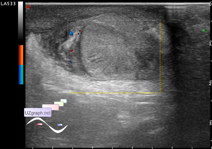
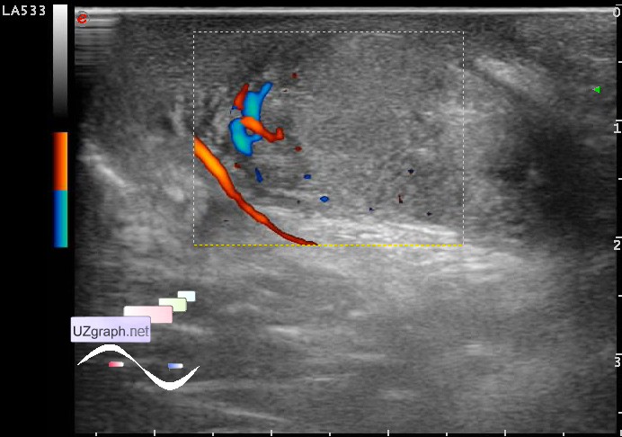
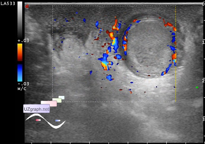
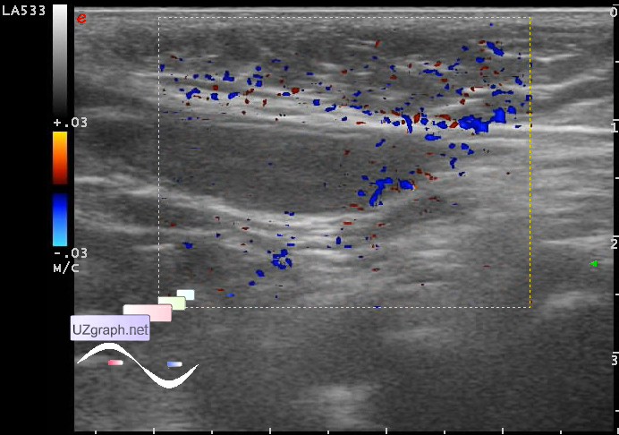
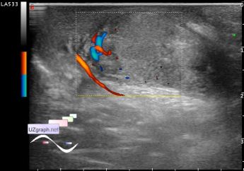
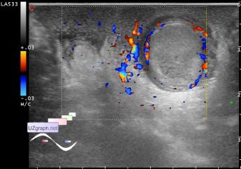
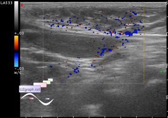
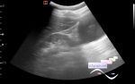
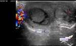
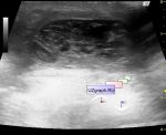
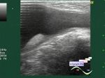
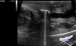
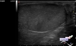
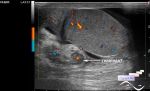
| Child 9 month old admitted to the reception department with a syndrome of scrotum edema, the surgeon sent for an ultrasound. On ultrasound the scrotum wall on both sides thickened, blood flow in the wall of the left scrotum greatly enhanced. The right testicle in the proximal third of the right inguinal canal size 21x7mm (cryptorchidism?), Blood flow in the CFM is not determined. The left testicle in the scrotum, the size 19x15 mm, round shape, inhomogeneous echostructure, hyperechoic with hypoechoic inclusions, in the CFM and DPD blood flow is visualized only in the head of epididymis and around the contour of the left testicle(so-called "Ring of Fire", the blood should go somewhere), in the spectral pulsed doppler mode in the head of epididymis determined only venous blood flow waveform, no arterial blood flow waveform is received, allegedly incomplete torsion of the left testicle. PS. Clear information on the anatomy textbook of M.Sapin, testicle and epididymis is supplying by blood from the testicular artery (a branch of the abdominal aorta) and partly from the artery of the vas(ductus) deferens (deferential artery, a branch of the internal iliac artery) which anastomoses with the testicular artery. However, it's an epididymis supplied with blood from two sources (which are there anastomose), ie from the testicular and deferential artery, and despite the fact that those arteries both are in the spermatic cord and can be torsed, but in the case of torsion is in the epididymis due to its double blood supply can be visualized blood flow and venous waveform of blood flow does not necessarily actually venous, it may well be poststenotic arterial blood flow. So in this case, torsed testicle, epididymis not completely torsed (still supplied with blood). Once I heard the surgeon's comment that if we see in the epididymis the blood flow, so there is no torsion, and it is certainly not right. Therefore, if you do not visualize blood flow in the testicle, but visualize it in the epididymis, you can not be satisfied because it's a torsion anyway! external link | |