Log in    RU  


Main page :: Forum :: Ultrasound cases - Sonograms, cine-loops etc. ::

Focal thyroiditis plus


Tags: Thyroid gland sonography, Esaote MyLab 70, Images, Video, Clinical report, Pediatric

   Posts
 
  Focal thyroiditis plus
#1

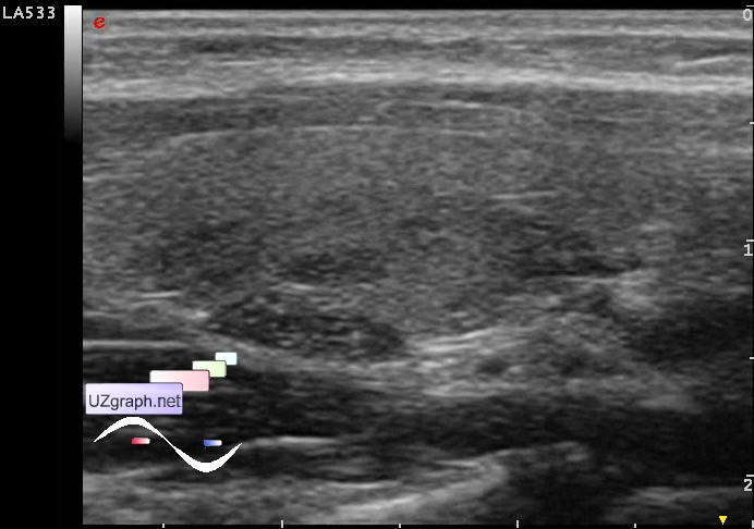
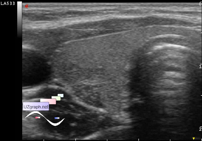
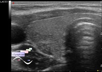
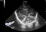
    A child of 9 years old came for a control thyroid ultrasound of the lesion up to 5 mm in one lobe, without any supporting documents.

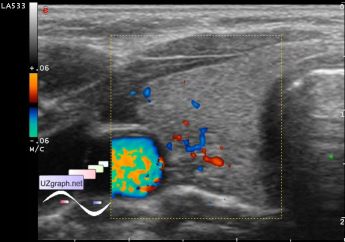
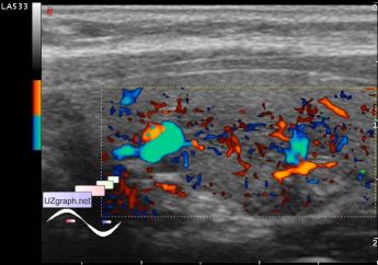
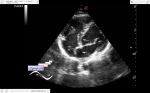
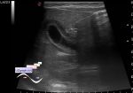
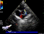
    At US in the posterior part of the right lobe of the thyroid gland is visualized the area of the heterogenicity type like isoechoic node, at CFM/DPD with increased blood flow, size of 19x9x5mm (thyroiditis? etc.?). Posterior to this area renders hypoechoic oval lesion with echo-structure similar to the thymus (thymus extra lobe? etc.?), size of 2x7mm.

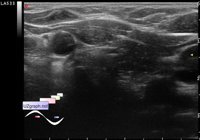
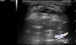
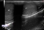
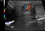
    PS. For comparison, 6 attachment - the thymus of the same child.

    external link

:: attachments(7) ::

:: file 1 ::

:: file 2 ::

:: file 3 ::

:: file 4 ::

:: file 5 ::

:: file 6 ::

:: file 7 ::


 Random 
Ultrasound cases - Twinkling artifact
    

Ultrasound cases - Beads eggs
    

Scientific - Playlist Pediatric sonography

Ultrasound cases - Medullary Rings
    

Ultrasound cases - Suspected intestinal pneumatosis
    

 Dictionary 
LVEDD

TV

BSA

LVESV

RVOT

Color M-mode

TI-RADS

TVUS

B-mode

EFW


 Calculators 
Body surface area

Teicholz systolic cardiac function

Gestational age by Ultrasound

Myocardial mass

TI-RADS


 Similar topics 
Ultrasound cases - Tirads 5 multinodular
    

Ultrasound cases - Pediatric Tirads 5
    

Ultrasound cases - Tirads 5
    

Ultrasound cases - Thyroiditis 2
    

Ultrasound cases - Diffuse-focal thyroid changes, parathyroid glands adenomas
    

Ultrasound cases - Accessory Thyroid Lobe
    

Ultrasound cases - Area with hyperechoic dot inclusions
    

Ultrasound cases - Thyroiditis
    

Ultrasound cases - Hypoplasia of the left thyroid lobe
    

Ultrasound cases - Cystic-solid volume of the thyroid gland