Log in    RU  


Main page :: Forum :: Ultrasound cases - Sonograms, cine-loops etc. ::

Extra flow in the heart


Tags: Cardiac sonography(Echocardiography), Medison Sonoace R7, Images, Video, Clinical report, Pediatric

   Posts
 
  Extra flow in the heart
#1

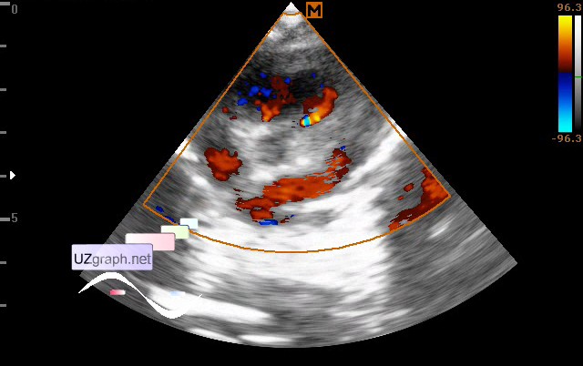
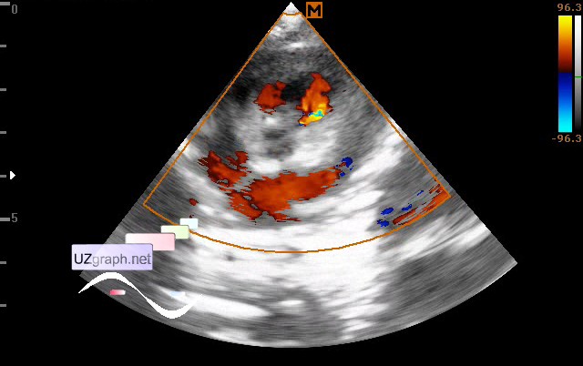
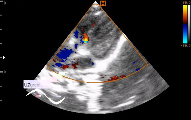
    Child 2 months. Came to the screening, before the problems with the heart was not revealed.

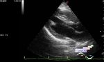
    At the CFM and PW: in the apical 4-chamber oblique view in the projection of the basal part of IVS, in the parasternal oblique view along the short axis in the area of the RVOT - additional flow are visualized. Flow up to 2-3 mm in the base with direction from the myocardium to the RVOT. V max. up to 1.5 m / s (slit-shaped IVSD? coronary fistula?).

    external link

:: attachments(4) ::

:: file 1 ::

:: file 2 ::

:: file 3 ::

:: file 4 ::


 Random 
Ultrasound cases - Inflamed mammary cyst(mastitis)
    

Ultrasound cases - Suspected liver hemangioma
    

Ultrasound cases - Cholelithiasis 3
    

Ultrasound cases - Increased periventricular echogenicity
    

Ultrasound cases - Diffuse changes of the testicle in a teenager (orchitis)
    

 Dictionary 
BSA

TI-RADS

TAMEAN

PW

MSD

PASP

Ved

LVEDD

PADP

HR


 Calculators 
Estimated fetal weight by Ultrasound

Resistive index

Gestational age by Ultrasound

Type of an infant hip joint

TI-RADS


 Similar topics 
Ultrasound cases - Mitral valve cyst
    

Ultrasound cases - Electrode in the right heart after installing of heart pacemaker
    

Ultrasound cases - Chylopericardium, lung atelectasis, cardiomegaly, ascites
    

Ultrasound cases - Asymmetric LVH, aortic regurgitation
    

Ultrasound cases - Coronary fistula from LCA?
    

Ultrasound cases - Floating IAS aneurysm
    

Ultrasound cases - Aortic Stenosis
    

Ultrasound cases - Floating Accessory Pulmonary Valve Tissue
    

Ultrasound cases - Hyperechoic linear structure in the left atrium
    

Ultrasound cases - Hydropericardium / tamponade