Log in    RU  


Main page :: Forum :: Ultrasound cases - Sonograms, cine-loops etc. ::

Discharge from the navel


Tags: Abdomen sonography, Urinary tract sonography, Esaote MyLab 70, Images, Video, Clinical report, Pediatric

   Posts
 
  Discharge from the navel
#1

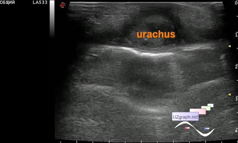
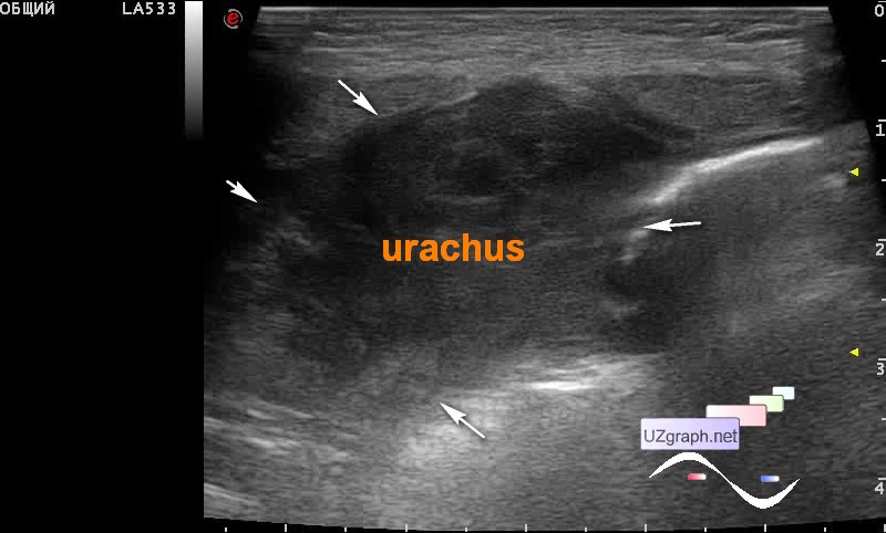
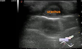
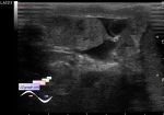
    An infant at the Children's City Clinical Hospital with a history of surgery to remove some kind of ovarian cyst (there was no documents about). At this moment she was admitted to the ED with complaints of discharge from the navel and with suspicion of a fistula was sent for an ultrasound scan.

    On ultrasound behind the bladder and above one of the ovaries there was a round lesion of a solid type with a cystic component (differential diagnosis: abscess, textiloma, etc.), on the CFM with blood flow, mainly around the lesion. From the navel to the bladder and the left lateral canal there was an another lesion of a heterogeneous structure (an approximate volume of up to 50 ml, differential diagnosis: urachus, abscess, etc.), pushing the intestines down and to the sides, when pressed by the probe, a long one curd consistency yellow "sausage" came out of the navel. It was not possible to exclude the relationship between these two lesions.

    external link

:: attachments(4) ::

:: file 1 ::

:: file 2 ::

:: file 3 ::

:: file 4 ::


 Random 
Ultrasound cases - Another GB polyp
    

Ultrasound cases - Tetralogy of Fallot
    

Ultrasound cases - GERD at WST
    

Ultrasound cases - Linea alba hernia vs lipoma part deux
    

Ultrasound cases - Diffuse-focal changes in the liver, thick left kidney
    

 Dictionary 
PG

PA

RVOT

CW

M-mode

CRL

Qp-Qs

FVI

GS

CFM


 Calculators 
Pressure gradient

Qp:Qs

Volume (ellipse formula)

Resistive index

Thyroid volume


 Similar topics 
Ultrasound cases - Portal hypertension
    

Ultrasound cases - Lesion in the projection of the adrenal gland 2
    

Ultrasound cases - Gallbladder, saw sign
    

Ultrasound cases - Ileotiphlitis and mesadenitis
    

Ultrasound cases - Cholelithiasis and hyperechoic gallbladder wall
    

Ultrasound cases - Portal vein spontaneous enhancement (mononucleosis)
    

Ultrasound cases - Hypoechoic spleen lesion
    

Ultrasound cases - Acute Appendicitis, periappendicular infiltrate
    

Ultrasound cases - Cavernous Transformation of the Portal Vein
    

Ultrasound cases - Diffuse-focal changes in the liver, thick left kidney