Log in    RU  


Main page :: Forum :: Ultrasound cases - Sonograms, cine-loops etc. ::

Neonatal hemorrhagic syndrome


Tags: Brain sonography(Neurosonography), Abdomen sonography, GE Logiq Book XP, Images, Video, Clinical report, Pediatric

   Posts
 
  Neonatal hemorrhagic syndrome
#1

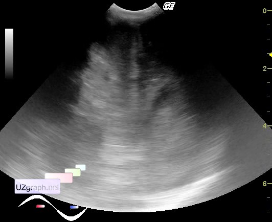
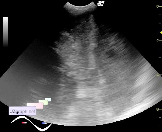
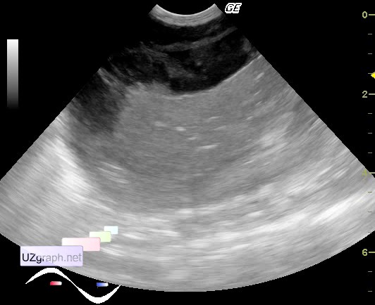
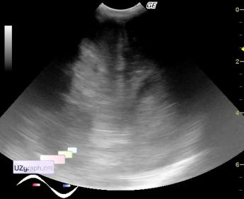
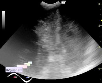
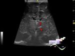
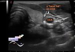
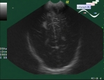
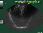
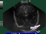
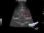
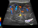
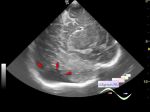
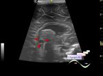
    Premature newborn in ICU, according to colleagues, was transferred from an infectious diseases hospital with suspected intestinal perforation.

    Visually, the right side of the head has an uneven surface, periodically with dark crimson crusts; 2/3 of the abdomen, on the right and down, is a rounded zone of elevation of a dark crimson and blue hue.

    On ultrasound, in the right posterior-lateral sections, there is an intracranial anechoic lesion approximately 4x3 cm in size, which displacing, compressing the right lateral ventricle to the center, presumably an intracranial(epidural?) hematoma (files 1-2, 5); In the abdominal cavity, in the projection of liver there is a heterogeneous lesion like a pocket of fluid (anechoic with multiple septa), presumably a hematoma (liver or abdominal cavity, files 3-5).

    On ultrasound of the kidneys, blood flow on both sides with a short diastolic phase and RI 0.8-0.9 (renal failure).

    external link

:: attachments(5) ::

:: file 1 ::

:: file 2 ::

:: file 3 ::

:: file 4 ::

:: file 5 ::


 Random 
Ultrasound cases - Coronary fistula from the left coronary artery
    

Ultrasound cases - Hypoechoic spleen lesion
    

Ultrasound cases - Wrist abscess
    

Ultrasound cases - Liver cirrhosis and thickened omentum
    

Ultrasound cases - Enlargement of the appendix
    

 Dictionary 
Anatomical M-mode

LMP

Aliasing doppler artifact

IVC

PSV

LVEDV

HR

MC

PADP

PD


 Calculators 
Volume (ellipse formula)

Estimated fetal weight by Ultrasound

TI-RADS

Resistive index

Pulmonary artery pressure


 Similar topics 
Ultrasound cases - Echogenic brain foci
    

Ultrasound cases - Subdural hemorrhage 2
    

Ultrasound cases - Willis circle at ultrasound
    

Ultrasound cases - Periventricular hyperechogenicity
    

Ultrasound cases - Combined (internal and external) hydrocephalus
    

Ultrasound cases - Subdural hemorrhage
    

Ultrasound cases - Cyst in the projection near the posterior horn of the lateral ventricle
    

Ultrasound cases - Subdural collection
    

Ultrasound cases - Anechogenic inclusion in the posterior cranial fossa
    

Ultrasound cases - Cyst of the base of the brain