Log in    RU  


Main page :: Forum :: Ultrasound cases - Sonograms, cine-loops etc. ::

Right Heart Enlargement


Tags: Cardiac sonography(Echocardiography), GE Logiq 400 MD, Clinical report, Pediatric

   Posts
 
  Right Heart Enlargement
#1

    A premature newborn was sent for a follow-up ultrasound of the heart (Echocardiography) to the public clinic, in the extract from the maternity hospital: PFO 2 mm, PDA, no dilatation of the heart chambers was noted.

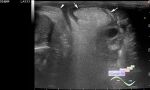
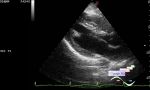
    On the current ultrasound of the heart, dilatation of the right heart is visualized with the prevalence (predominance) of the right heart over the left (i.e. the right chambers are wider than the left; differential diagnosis: severe pulmonary hypertension, HLHS, etc.), blood shunting is also visualized from the left to the right through an ASD of the ostium secundum type up to 6 mm in size with Vmax up to 1 m/s. Vmax in PA up to 1.7 m/s.


 Random 
Ultrasound cases - ADPKD
    

Ultrasound cases - Indirect inguinal hernia in a girl
    

Ultrasound cases - Lenticulostriate vasculopathy
    

Ultrasound cases - Pneumonia
    

Scientific - Playlist Vascular sonography

 Dictionary 
BPD

FNA

RAP

Doppler angle

VTI

TV

Ved

LN

PRF

LVEDD


 Calculators 
Qp:Qs

Myocardial mass

Gestational age by Ultrasound

Volume (ellipse formula)

Teicholz systolic cardiac function


 Similar topics 
Ultrasound cases - Bicuspid aortic valve 2
    

Ultrasound cases - Mitral valve cyst
    

Ultrasound cases - Electrode in the right heart after installing of heart pacemaker
    

Ultrasound cases - Chylopericardium, lung atelectasis, cardiomegaly, ascites
    

Ultrasound cases - Asymmetric LVH, aortic regurgitation
    

Ultrasound cases - Coronary fistula from LCA?
    

Ultrasound cases - Floating IAS aneurysm
    

Ultrasound cases - Aortic Stenosis
    

Ultrasound cases - Floating Accessory Pulmonary Valve Tissue
    

Ultrasound cases - Hyperechoic linear structure in the left atrium