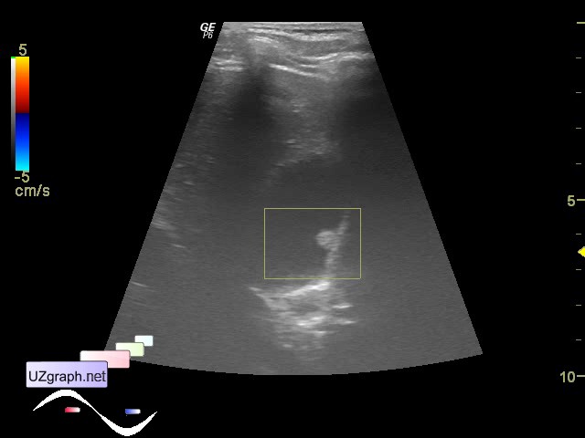
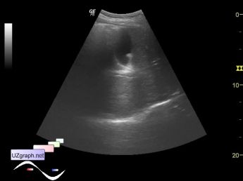
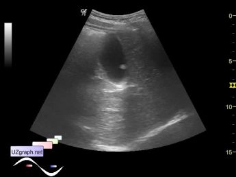
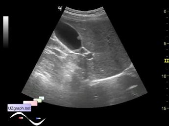
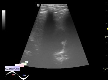
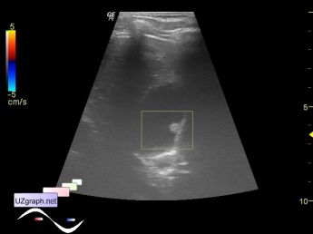
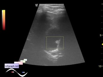
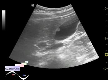
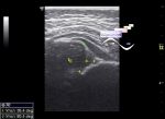
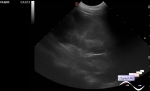
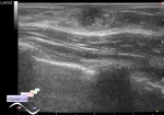
Log in    RU  


Main page :: Forum :: Ultrasound cases - Sonograms, cine-loops etc. ::

Polyp of the gallbladder


Tags: Abdomen sonography, Images, Video, Clinical report, GE Logiq P6, Pediatric

   Posts
 
  Polyp of the gallbladder
#1

    Teenager on medical examination.

    In the projection of the front wall of the gallbladder there is a rounded mass with solid structure, up to 5 mm, without blood flow at CFM and PD, does not move in a standing position (attachment 7), presumably polyp.

:: attachments(8) ::

:: file 1 ::

:: file 2 ::

:: file 3 ::

:: file 4 ::

:: file 5 ::

:: file 6 ::

:: file 7 ::

:: file 8 ::


 Random 
Ultrasound cases - Ovarian apoplexy 3
    

Ultrasound cases - Bifurcated rib (Lushka's fork)
    

Ultrasound cases - Colloid nodules
    

Ultrasound cases - PDA before surgery
    

Ultrasound cases - Condition after appendectomy
    

 Ad 


 Dictionary 
LVPWd

PA

PD

PSAX

GA

ESV

PRF

LVLs

Anatomical M-mode

LAP


 Calculators 
"Bullet" formula (1 disk) ejection fraction

Modified Simpson’s Rule formula (3 disks) ejection fraction

TI-RADS ACR

Pulmonary artery pressure

Volume (ellipse formula)


 Similar topics 
Ultrasound cases - Hirschsprung disease on ultrasound
    

Ultrasound cases - Intestinal Infiltrate
    

Ultrasound cases - Abdominal cryptorchidism
    

Ultrasound cases - Diffuse heterogeneous liver
    

Ultrasound cases - Postoperative abdominal wall fistula
    

Ultrasound cases - Stump appendicitis
    

Ultrasound cases - Liver scar after resection
    

Ultrasound cases - Subdiaphragmatic movable fluid collection (UFO)
    

Ultrasound cases - Lesion of the spleen or the case when it is easy to not notice
    

Ultrasound cases - I’m normal but others ...