Log in    RU  


Main page :: Forum :: Ultrasound cases - Sonograms, cine-loops etc. ::

Linea alba hernia vs lipoma part deux


Tags: Soft tissues sonography, Abdomen sonography, Images, Video, Clinical report, Esaote MyLab 70, Pediatric

   Posts
 
  Linea alba hernia vs lipoma part de...
#1

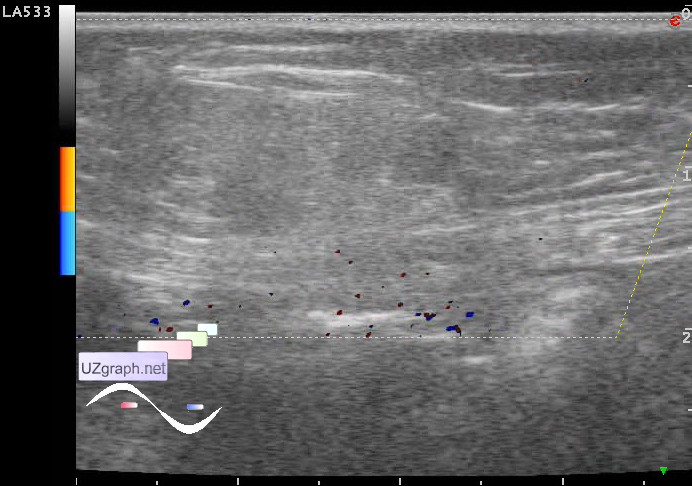
    A child 5 years old, with the lesion of the anterior abdominal wall after the surgeon consultation is aimed at US with suspected anterior abdominal wall lipoma.

    Visually in the upper abdomen slightly to the side of the midline slightly noticeable bulging of the skin. Parents denied the trauma.

    On US there is a subcutaneous oval shape lesion, about 2 cm in size, adjacent to the rectus abdominis muscle and the linea alba, has an unclear contours, heterogeneous an / iso echogenic structure (fluid?), an acoustic shadow / defect of linea alba in one of the poles, on DPD(CFM) without blood flow.

    Presumably hernia of the linea alba with a greater omentum.

    external link

:: attachments(6) ::

:: file 1 ::

:: file 2 ::

:: file 3 ::

:: file 4 ::

:: file 5 ::

:: file 6 ::


 Random 
Ultrasound cases - Polyps of the gallbladder
    

Ultrasound cases - Hydronephrosis of half of horseshoe kidney
    

Ultrasound cases - Infrapatellar bursitis
    

Ultrasound cases - Double inferior vena cava
    

Ultrasound cases - Subdural hemorrhage 2
    

 Dictionary 
BPD

PVR

M-mode

B-mode

CRL

PA

RVOT

GA

ESV

MC


 Calculators 
Body surface area

Gestational age by Ultrasound

Myocardial mass

Teicholz systolic cardiac function

Volume (ellipse formula)


 Similar topics 
Ultrasound cases - Penetrating injury of the femur
    

Ultrasound cases - Cephalohematoma
    

Ultrasound cases - Urachus fistula
    

Ultrasound cases - Cranial mass
    

Ultrasound cases - Pilonidal coccyx cyst
    

Ultrasound cases - Palm fistula, in search of pencil lead
    

Ultrasound cases - Postoperative abdominal wall fistula
    

Ultrasound cases - Nucks cyst
    

Ultrasound cases - Paraproctitis
    

Ultrasound cases - Submandibular area lesion