Log in    RU  


Main page :: Forum :: Ultrasound cases - Sonograms, cine-loops etc. ::

Osgood-Schlatter disease, tendinitis


Tags: Musculoskeletal sonography, Images, Video, Clinical report, Esaote MyLab 70, Pediatric

   Posts
 
  Osgood-Schlatter disease, tendiniti...
#1

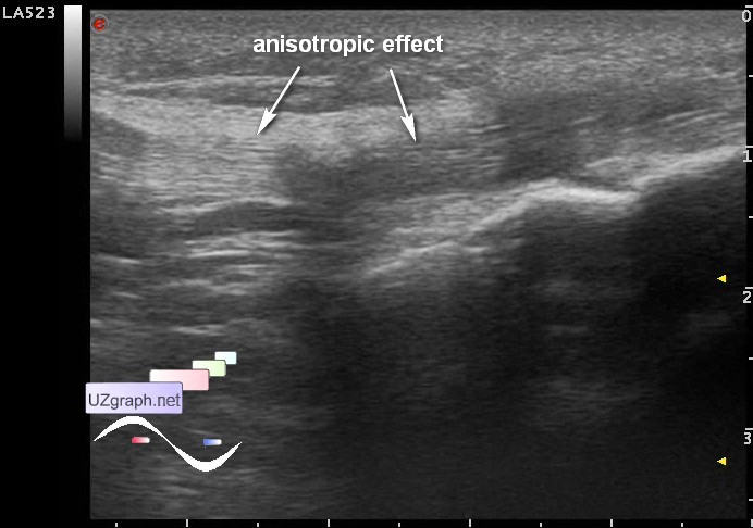
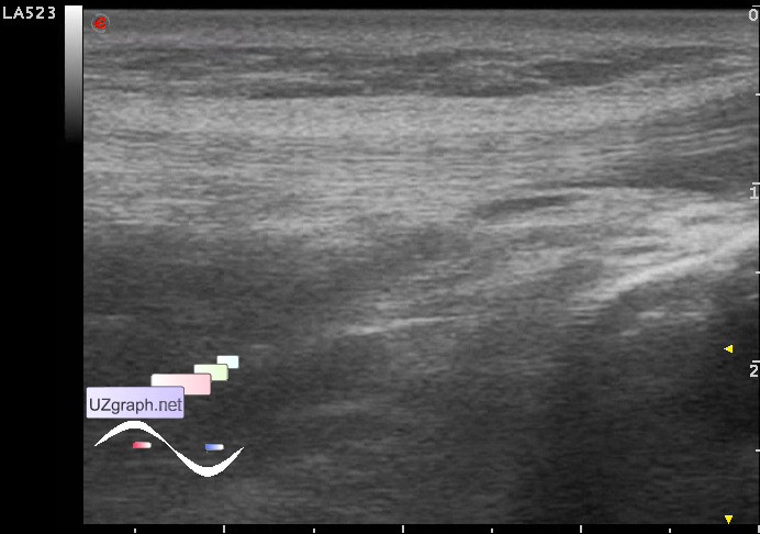
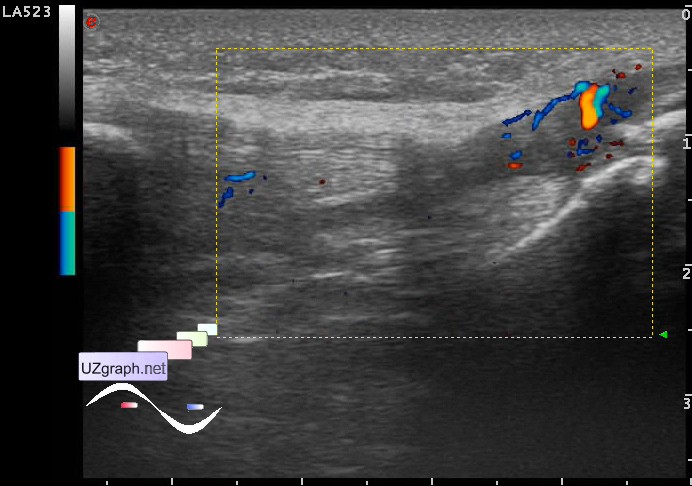
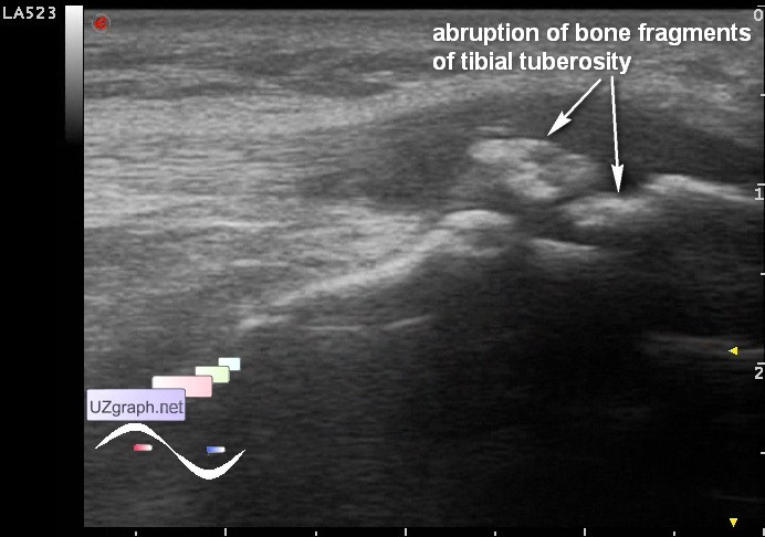
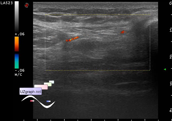
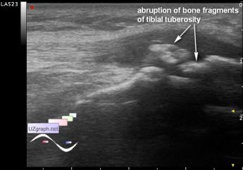
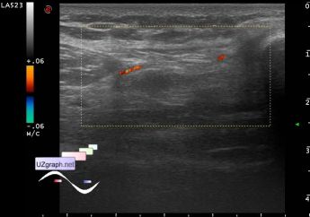
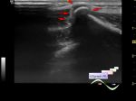
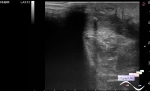
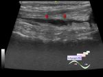
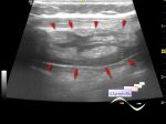
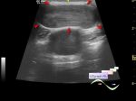
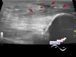
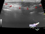
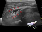
    A teenager with a history of recurrent combined injuries of the one knee, by his words: first he is fell, a few days after he got a strike and there is something out of his knee was jumping... in emergency room he had a Roentgen with diagnosis of Osgood-Schlatter disease, but orthopedist asked to see a ligaments condition on ultrasound...

    At US in the projection of the distal part of the patellar tendon in the rectified knee position visualized hypoechoic area and even some impression (looks like a partial rupture), but in the bent knee position the ligament becomes uniform echogenicity (anisotropic effect - an ultrasound artifact) and flat shapes. On the DPD in this area visualized increased blood flow (tendonitis of the patellar tendon) and bone fragments of tibial tuberosity (Osgood-Schlatter disease) . Also in the projection of quadriceps tendon on the DPD visualized a small increase in blood flow (tendinitis of the quadriceps tendon), compared with blood flow in the projection of the patellar tendon. In a healthy knee blood flow in these areas, at the same Doppler settings, was not visualized.

    Files:

    1 anisotropic effect of patellar tendon in the rectified knee position

    2 almost complete disappearance of this effect when bending the knee

    3 DPD - increased blood flow in the distal patellar tendon

    4 abruption of bone fragments of tibial tuberosity

    5 DPD - increased blood flow in the quadriceps tendon

    external link

:: attachments(6) ::

:: file 1 ::

:: file 2 ::

:: file 3 ::

:: file 4 ::

:: file 5 ::

:: file 6 ::


 Random 
Ultrasound cases - Conglomerate in the right iliac area
    

Ultrasound cases - Varicocele
    

Ultrasound cases - Hemorrhagic ovarian cyst
    

On-call room - A new method of labor induction ... joke

Ultrasound cases - Scapula exostosis
    

 Dictionary 
PD

TAV

LVPWd

SF

LVESV

DPD

MSD

LVOT

EFW

TI-RADS


 Calculators 
Resistive index

Volume (ellipse formula)

Type of an infant hip joint

Myocardial mass

Estimated fetal weight by Ultrasound


 Similar topics 
Ultrasound cases - Penetrating injury of the femur
    

Ultrasound cases - Cephalohematoma
    

Ultrasound cases - Cranial mass
    

Ultrasound cases - Recurrent shin trauma
    

Ultrasound cases - Knee joint synovitis
    

Ultrasound cases - Thickening and heterogeneity of the intercostal space soft tissues
    

Ultrasound cases - Chest wall lesion
    

Ultrasound cases - Hockey Elbow
    

Ultrasound cases - Combination of left shoulder lesions
    

Ultrasound cases - Baker's cyst and intramuscular hematoma