Log in    RU  


Main page :: Forum :: Ultrasound cases - Sonograms, cine-loops etc. ::

Nephroptosis


Tags: Urinary tract sonography, Images, Video, Clinical report, Esaote MyLab 70, Pediatric

   Posts
 
  Nephroptosis
#1

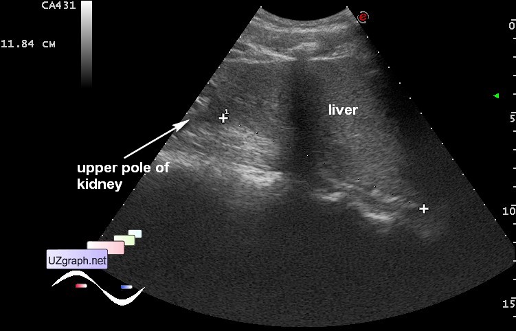
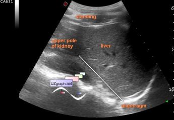
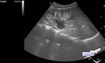
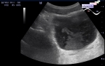
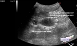
    Teenager with periodically pain in the right low abdomen quadrant, suspected acute appendicitis was addressed to ultrasound.

    No sign of appendicitis there was found but right kidney has a high mobility - going down about 7 cm in vertical body position.

    Picture demonstrate a measurement of distance between diaphragm and upper pole of right kidney, which was 5 cm horizontal body position(all right kidney was seen), and become 12 cm in vertical.

:: attachments(2) ::

:: file 1 ::

:: file 2 ::

  00:35 29-11-2015
  Another case
#2

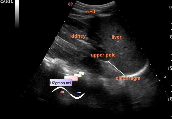
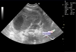
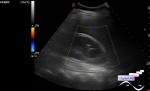
    Teenager 17 years-old with flank pain and right kidney nephroptosis.

    At US from the side body access: the distance between the upper pole of the kidneys and the diaphragm in the supine position is 3 cm and 7.5 cm in standing position.

:: attachments(2) ::

:: file 1 ::

:: file 2 ::


 Random 
Ultrasound cases - Inguino-scrotal hernia and enlarged appendix
    

Ultrasound cases - Urachal sinus
    

Ultrasound cases - Combined (internal and external) hydrocephalus
    

Ultrasound cases - Biceps rupture
    

Ultrasound cases - "Sand" in the kidneys
    

 Dictionary 
HR

Doppler angle

TAMEAN

LAP

FNA

SF

PA

MSD

TV

Ved


 Calculators 
Thyroid volume

Teicholz systolic cardiac function

TI-RADS

Myocardial mass

Pressure gradient


 Similar topics 
Ultrasound cases - Autosomal recessive polycystic kidney disease (ARPKD)
    

Ultrasound cases - Bladder sludge
    

Ultrasound cases - Bright kidneys
    

Ultrasound cases - Ileostoma stenosis and nephropathy
    

Ultrasound cases - Renal pelvis clot
    

Ultrasound cases - Renal pelvis valve-stone
    

Ultrasound cases - Condition after treatment of a kidney cyst
    

Ultrasound cases - Hydronephrotic transformation & polypous bladder wall
    

Ultrasound cases - Pyelitis, cystitis
    

Ultrasound cases - Lesion in the projection of the adrenal gland 2