Log in    RU  


Main page :: Forum :: Ultrasound cases - Sonograms, cine-loops etc. ::

Smart alien


Tags: Gastrointestinal sonography, GE Vivid 3, Images, Video, Clinical report, Adult

   Posts
 
  Smart alien
#1

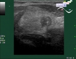
    The patient 18 years old without complaints came on the abdomen ultrasound because of the prophylactic medical examination program.

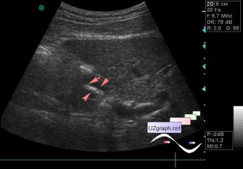
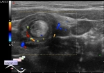
    On ultrasound in the projection of the lumen of the dilated segment of the duodenum, a mobile hyperechoic tubular structure up to 4 mm in diameter (ascaride?) is visualized.

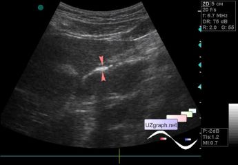
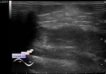
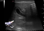
    At the end of the study, I decided to re-examine the zone of interest and to my surprise I did not immediately notice where this structure disappeared (fig. 2), but she decided to lie down along the wall of the intestine (a smart move to merge with the landscape).

:: attachments(3) ::

:: file 1 ::

:: file 2 ::

:: file 3 ::


 Random 
Ultrasound cases - Testicular torsion after surgery
    

Ultrasound cases - Thelarche
    

Ultrasound cases - MV fistula?
    

Ultrasound cases - Isolated spleen calcification
    

Ultrasound cases - Clinically suspected appendicitis 3
    

 Dictionary 
CRL

GA

Doppler angle

LVESV

Freeze

SV

SF

TV

TAV

FNA


 Calculators 
Type of an infant hip joint

Gestational age by Ultrasound

Estimated fetal weight by Ultrasound

Teicholz systolic cardiac function

TI-RADS


 Similar topics 
Ultrasound cases - Hirschsprung disease on ultrasound
    

Ultrasound cases - Intestinal Infiltrate
    

Ultrasound cases - Subhepatic appendicitis and omentitis
    

Ultrasound cases - Diffuse heterogeneous liver
    

Ultrasound cases - Stump appendicitis
    

Ultrasound cases - Ileostoma stenosis and nephropathy
    

Ultrasound cases - Periappendicular infiltrate
    

Ultrasound cases - Intestinal obstruction, GB wall thickening
    

Ultrasound cases - Ileotiphlitis and mesadenitis
    

Ultrasound cases - Acute Appendicitis, periappendicular infiltrate