Log in    RU  


Main page :: Forum :: Ultrasound cases - Sonograms, cine-loops etc. ::

Megacolon


Tags: Abdomen sonography, Gastrointestinal sonography, GE Logiq E, Clinical report, Pediatric

   Posts
 
  Megacolon
#1

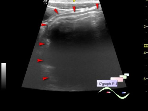
    A 3-year-old child was sent for an abdominal ultrasound to a public clinic with complaints of rare stools, as it became known after an ultrasound scan, the child had previously undergone an X-ray contrast study of the large intestine in the hospital with a diagnosis of megacolon.

    On ultrasound in the projection of the left hypo- / mesogastrium, a section of the large intestine enlarged to about 5.4 cm, presumably the sigmoid colon, is visualized (diff. diagnosis: Hirschsprung's disease, dolichosigma, etc.).

    A consultation with a gastroenterologist is recommended.


    external link


 Random 
Ultrasound cases - Pseudotumor of the liver
    

Ultrasound cases - Hydropericardium / tamponade
    

Ultrasound cases - PDA forth and back
    

Ultrasound cases - Goiter Hashimoto
    

Ultrasound cases - Cholelithiasis plus
    

 Dictionary 
SF

LVEDD

ESD

B-mode

BSA

Qp-Qs

LVPWd

LMP

PSV

EDD


 Calculators 
Body surface area

Gestational age by Ultrasound

Estimated fetal weight by Ultrasound

Thyroid volume

Type of an infant hip joint


 Similar topics 
Ultrasound cases - Hirschsprung disease on ultrasound
    

Ultrasound cases - Intestinal Infiltrate
    

Ultrasound cases - Abdominal cryptorchidism
    

Ultrasound cases - Diffuse heterogeneous liver
    

Ultrasound cases - Postoperative abdominal wall fistula
    

Ultrasound cases - Stump appendicitis
    

Ultrasound cases - Liver scar after resection
    

Ultrasound cases - Subdiaphragmatic movable fluid collection (UFO)
    

Ultrasound cases - Lesion of the spleen or the case when it is easy to not notice
    

Ultrasound cases - I’m normal but others ...