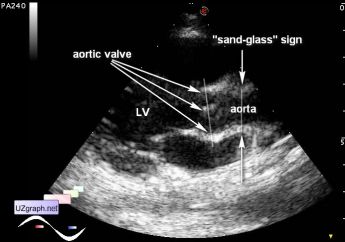
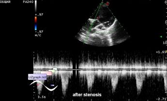
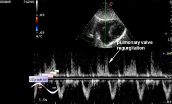
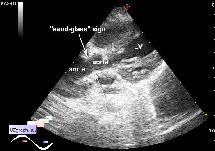
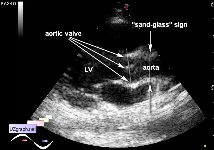
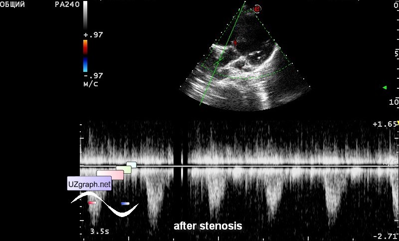
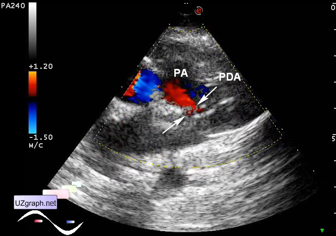
Supravalvular aortic stenosis
Tags: Cardiac sonography(Echocardiography), Esaote MyLab 70, Images, Video, Clinical report, Pediatric
| Posts | |||
| Supravalvular aortic stenosis | #1 |
| |||||
:: file 1 ::
 :: file 2 ::
 :: file 3 ::
 :: file 4 ::
 :: file 5 ::
 :: file 6 ::
 :: file 7 ::
 :: file 8 ::
 :: file 9 :: | |||||