Log in    RU  


Main page :: Forum :: Ultrasound cases - Sonograms, cine-loops etc. ::

Neck fistulas


Tags: Soft tissues sonography, Chest sonography, Images, Video, Clinical report, Esaote MyLab 70, Pediatric

   Posts
 
  Neck fistulas
#1

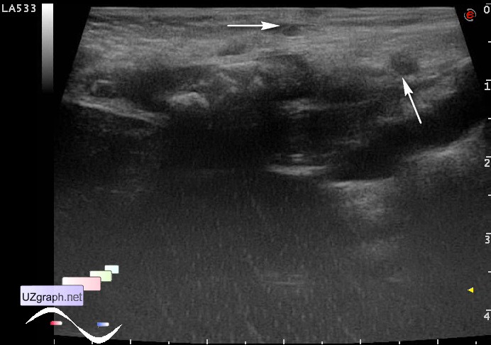
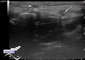
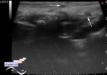
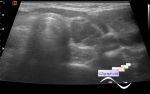
    Child 1 year old with suspected fistula / lateral neck cyst is aimed at ultrasound. Previously, child has already an abscess in the same location, which was drained. At this moment, parents noticed new spots.

    Visually, on the border of the neck and chest (in the area of left clavicle) 2 pink spots.

    At ultrasound of this area visualized 1-2(?) subcutaneous thin up to 2 mm in diameter curved channels - fistulas (branchial cleft fistulas?).

    external link

:: attachments(3) ::

:: file 1 ::

:: file 2 ::

:: file 3 ::


 Random 
Ultrasound cases - Cryptorchidism, microlithiasis, bladder lesion
    

Ultrasound cases - Periappendicular infiltrate
    

Ultrasound cases - Perimedullary rings sign
    

Ultrasound cases - Group of hepatic cysts
    

Ultrasound cases - Lateral neck cyst/ branchial cleft cyst
    

 Dictionary 
TVUS

Qp-Qs

LMP

PA

PW

PADP

TAV

TAMEAN

FVI

LVPWd


 Calculators 
Resistive index

Myocardial mass

Thyroid volume

Volume (ellipse formula)

Pressure gradient


 Similar topics 
Ultrasound cases - Penetrating injury of the femur
    

Ultrasound cases - Cephalohematoma
    

Ultrasound cases - Urachus fistula
    

Ultrasound cases - Cranial mass
    

Ultrasound cases - Pilonidal coccyx cyst
    

Ultrasound cases - Palm fistula, in search of pencil lead
    

Ultrasound cases - Postoperative abdominal wall fistula
    

Ultrasound cases - Nucks cyst
    

Ultrasound cases - Paraproctitis
    

Ultrasound cases - Submandibular area lesion