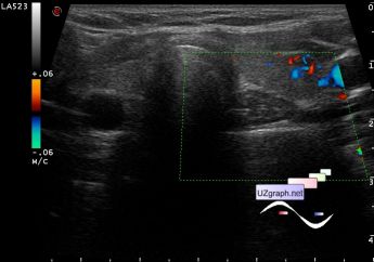
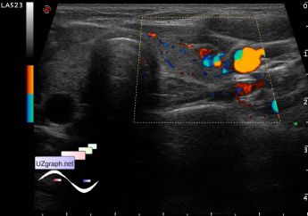
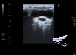
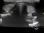
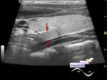
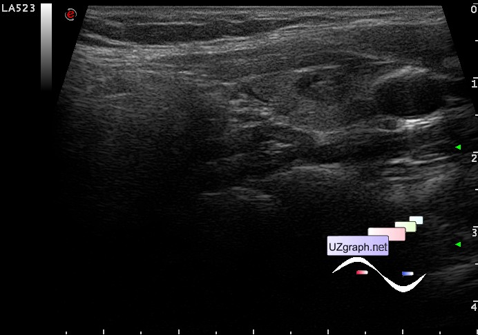
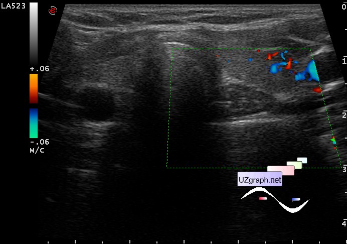
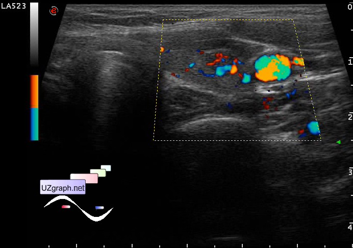
Richly vascularized thyroid nodule
Tags: Thyroid gland sonography, Images, Video, Clinical report, Esaote MyLab 70, Pediatric
| Posts | |||
| Richly vascularized thyroid nodule | #1 |
| |||||
:: file 1 ::
:: file 2 ::
 :: file 3 ::
 :: file 4 ::
 :: file 5 ::
 :: file 6 :: | |||||