Log in    RU  


Main page :: Forum :: Ultrasound cases - Sonograms, cine-loops etc. ::

Shoulder hemangioma


Tags: Soft tissues sonography, Images, Video, Clinical report, Esaote MyLab 70, Pediatric

   Posts
 
  Shoulder hemangioma
#1

    Child 5 months old with the shoulder lesion, close to the elbow, aimed to ultrasound from surgeon with diagnosis of hemangioma ...

    Visually proximal to elbow there is a slight change in color of the skin looks as birthmark up to 3mm around which there is a visible area of blueness up to 1 cm.

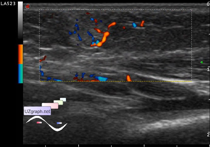
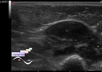
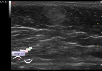
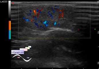
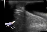
    On US subcutaneously visualized hyperechoic lesion 1,5x0,8 cm with an unclear border tending to the oval shape on CFM(DPD) is richly supplied with blood.

    external link

:: attachments(5) ::

:: file 1 ::

:: file 2 ::

:: file 3 ::

:: file 4 ::

:: file 5 ::


 Random 
Ultrasound cases - Ileotiphlitis and mesadenitis
    

Ultrasound cases - Neonatal breast enlargement
    

Ultrasound cases - Muscular IVSD
    

Ultrasound cases - I am an intelligent man but sometimes ...
    

Ultrasound cases - Cryptorchidism
    

 Dictionary 
EDD

B-mode

MC

PADP

PASP

HC

Gain

EDV

CO

BSA


 Calculators 
Estimated fetal weight by Ultrasound

Qp:Qs

Teicholz systolic cardiac function

Myocardial mass

Pulmonary artery pressure


 Similar topics 
Ultrasound cases - Penetrating injury of the femur
    

Ultrasound cases - Cephalohematoma
    

Ultrasound cases - Urachus fistula
    

Ultrasound cases - Cranial mass
    

Ultrasound cases - Pilonidal coccyx cyst
    

Ultrasound cases - Palm fistula, in search of pencil lead
    

Ultrasound cases - Postoperative abdominal wall fistula
    

Ultrasound cases - Nucks cyst
    

Ultrasound cases - Paraproctitis
    

Ultrasound cases - Submandibular area lesion