Log in    RU  


Main page :: Forum :: Ultrasound cases - Sonograms, cine-loops etc. ::

Frontal area lesion


Tags: Soft tissues sonography, Musculoskeletal sonography, Images, Video, Clinical report, Esaote MyLab 70, Pediatric

   Posts
 
  Frontal area lesion
#1

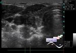
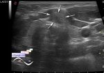
    Child 1 month old with lesion in the frontal area of the head after the surgeon's consultation with the diagnosis of fibroma.

    Visually on a side surface of frontal area there is a subcutaneous lesion up to 5 mm. By the words of the parents it became smaller now!

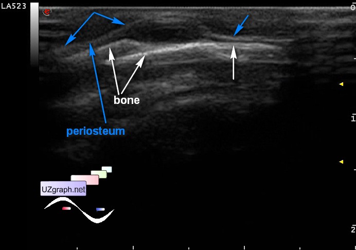
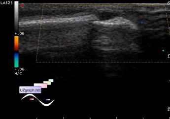
    At US in this projection there is a lesion up to 6 mm, an / hypoechoic, oval-shaped, projecting deeper to periosteum (cephalhematoma?), Without blood flow on CFM.

    external link

:: attachments(4) ::

:: file 1 ::

:: file 2 ::

:: file 3 ::

:: file 4 ::


 Random 
Ultrasound cases - Penetrating injury of the femur
    

Ultrasound cases - Left ventricular non-compaction
    

Ultrasound cases - Inguino-scrotal hernia and enlarged appendix
    

Ultrasound cases - Vaccination schedule 2
    

Ultrasound cases - Cyst of the epididymis and appendages
    

 Dictionary 
Color M-mode

PG

RVOT

AC

MC

PD

BSA

LVPWd

Anatomical M-mode

TV


 Calculators 
Myocardial mass

TI-RADS

Type of an infant hip joint

Volume (ellipse formula)

Pressure gradient


 Similar topics 
Ultrasound cases - Penetrating injury of the femur
    

Ultrasound cases - Cephalohematoma
    

Ultrasound cases - Urachus fistula
    

Ultrasound cases - Cranial mass
    

Ultrasound cases - Pilonidal coccyx cyst
    

Ultrasound cases - Palm fistula, in search of pencil lead
    

Ultrasound cases - Postoperative abdominal wall fistula
    

Ultrasound cases - Nucks cyst
    

Ultrasound cases - Paraproctitis
    

Ultrasound cases - Submandibular area lesion