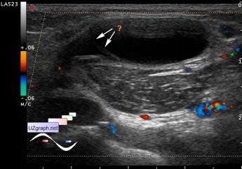
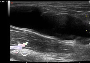
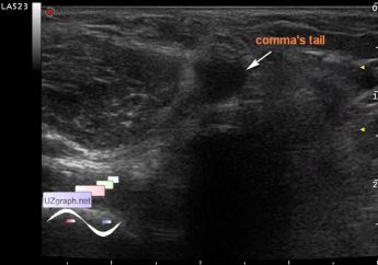
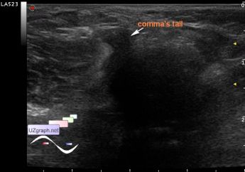
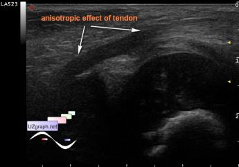
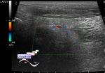
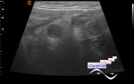
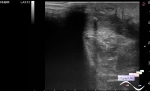
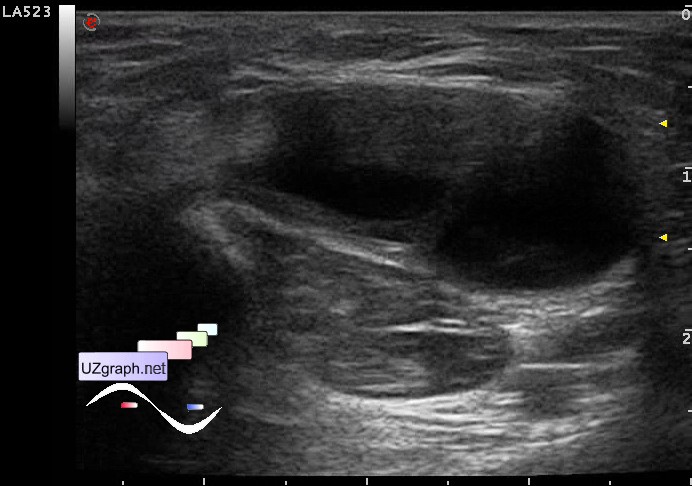
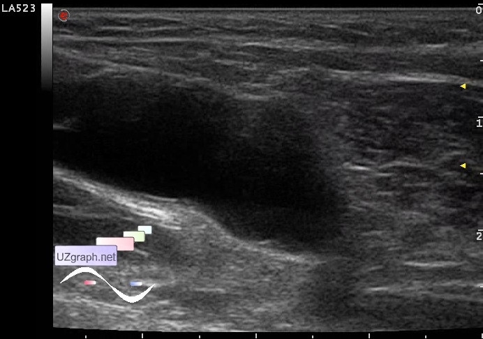
Classical Baker's cyst and the complexity ...
Tags: Musculoskeletal sonography, Soft tissues sonography, Images, Video, Clinical report, Esaote MyLab 70, Pediatric
| Posts | |||
| Classical Baker's cyst and the ... | #1 |
| |||||
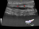
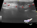
:: file 1 ::
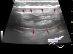
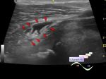
 :: file 2 ::
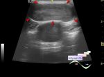
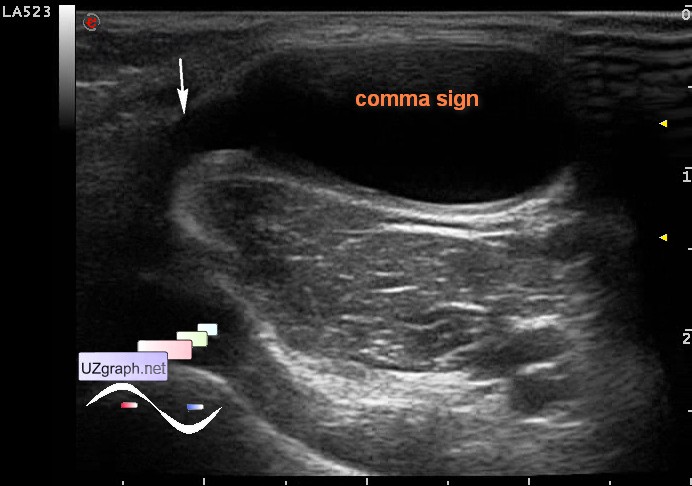
 :: file 3 ::
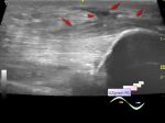
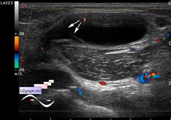
 :: file 4 ::
 :: file 5 ::
 :: file 6 ::
 :: file 7 ::
 :: file 8 ::
 :: file 9 ::
 :: file 10 :: | |||||