Log in    RU  


Main page :: Forum :: Ultrasound cases - Sonograms, cine-loops etc. ::

Polysplenia (heterotaxy, left isomerism)


Tags: Abdomen sonography, Esaote MyLab 70, Images, Video, Clinical report, Pediatric

   Posts
 
  Polysplenia (heterotaxy, left isome...
#1

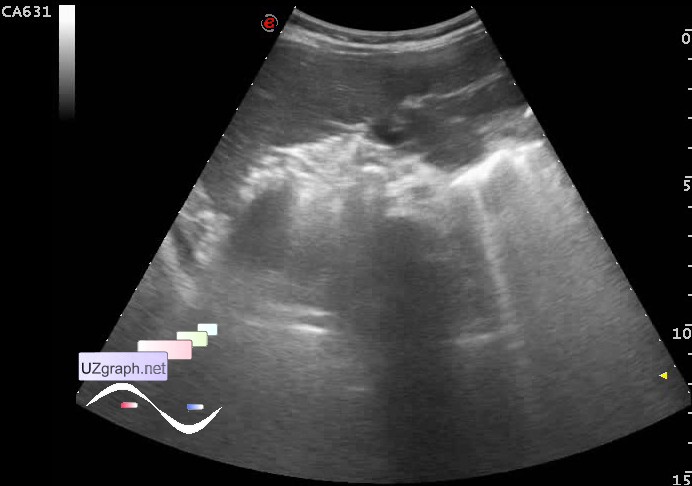
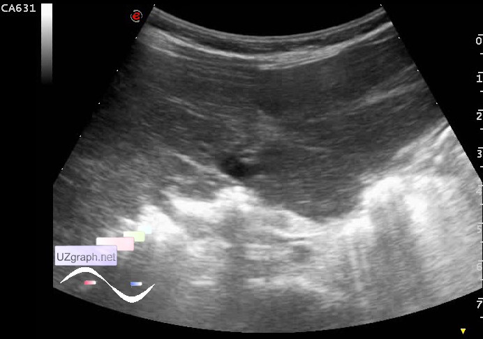
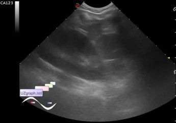
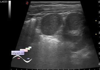
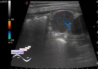
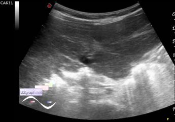
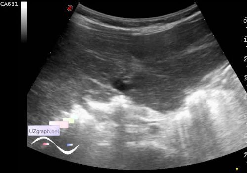
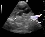
    Child of 10 months-old aims to control ultrasonography of abdomen, kidneys and heart with diagnoses by results of US at the age of 1 month: deformation of GB, pielectasis and PFO. Spleen described as normal.

    In the current US PFO and pielectasis not found, but found no spleen, in the form as we used to see it - it is represented by 5 or more isolated lobes rounded / oval shape a maximum size about of 3 cm, also of 2 cm and 1 cm. (polysplenia)

    Caudate lobe of liver extends beyond the border of the liver (enlarged?)

    The pancreas is not reliably visualized (shielded by intestinal gas? anomaly?). The left kidney is enlarged.

    Recommended CT of abdomen.

    Reference. Many of us are familiar with the term accessory spleen, they can be even two or more. Polysplenia term means not only having multiple spleens, but the absence of the main/parent spleen! This syndrome may be associated with a large range of other abnormalities of organs and systems. (Heterotaxy. Left isomerism)

    Read also. external link

    external link

:: attachments(6) ::

:: file 1 ::

:: file 2 ::

:: file 3 ::

:: file 4 ::

:: file 5 ::

:: file 6 ::

  10:58 21-05-2019
 
#2


    Possible variant of Riedel' s lobe

    external link


 Random 
Ultrasound cases - Varicocele
    

Ultrasound cases - Knee cyst
    

Ultrasound cases - Bicuspid aortic valve 2
    

Ultrasound cases - Goiter Hashimoto
    

Admin room - Mobile application for android
    

 Dictionary 
Freeze

EDD

LVESV

Qp-Qs

TAMEAN

M-mode

AC

TAV

PASP

MSD


 Calculators 
Estimated fetal weight by Ultrasound

Body surface area

Pulmonary artery pressure

Resistive index

Pressure gradient


 Similar topics 
Ultrasound cases - Hirschsprung disease on ultrasound
    

Ultrasound cases - Intestinal Infiltrate
    

Ultrasound cases - Abdominal cryptorchidism
    

Ultrasound cases - Diffuse heterogeneous liver
    

Ultrasound cases - Postoperative abdominal wall fistula
    

Ultrasound cases - Stump appendicitis
    

Ultrasound cases - Liver scar after resection
    

Ultrasound cases - Subdiaphragmatic movable fluid collection (UFO)
    

Ultrasound cases - Lesion of the spleen or the case when it is easy to not notice
    

Ultrasound cases - I’m normal but others ...