Log in    RU  


Main page :: Forum :: Ultrasound cases - Sonograms, cine-loops etc. ::

Abdominal syndrome & bladder diverticulum


Tags: Abdomen sonography, Urinary tract sonography, Gastrointestinal sonography, Esaote MyLab 70, Medison Sonoace R7, Images, Video, Clinical report, Pediatric

   Posts
 
  Abdominal syndrome & bladder divert...
#1

    A child of 8 years old entered in emergency department with suspected appendicitis, previously identified bladder diverticulum.

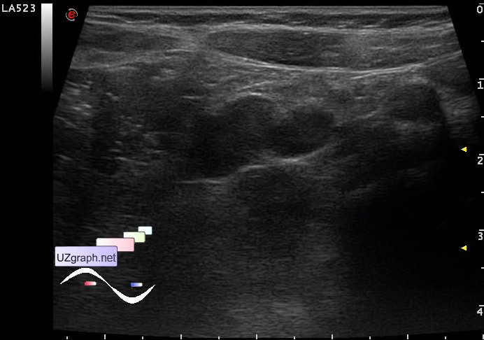
    In mesogaster visualized lymph nodes more than 5 to 12 mm (mesadenitis?)

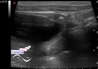
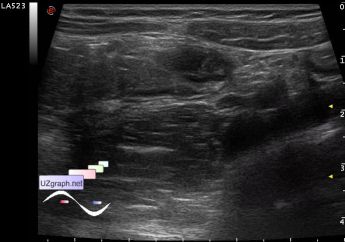
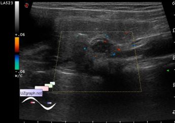
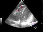
    In the right iliac region is rendered tubular structure up to 6.5 mm in diameter, with blood flow at CFM, rigid while probe compression, painful reaction when compression is not obtained (acute appendicitis?).

    In the right iliac region also is visualized with a thickened segment of the bowel, wall to 4mm (enterocolitis?)

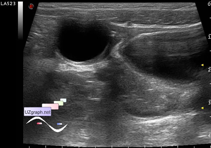
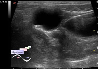
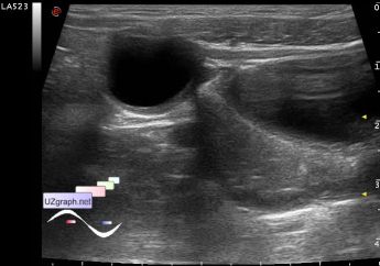
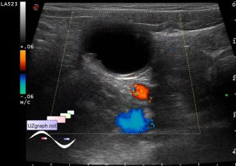
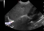
    The bladder is filled, right from the bladder visualized anechoic lesion oval shaped size of 1,5x2cm without blood flow at CFM, visually linking with the bladder thru channel to 2 mm in diameter (bladder diverticulum).

    external link

:: attachments(11) ::

:: file 1 ::

:: file 2 ::

:: file 3 ::

:: file 4 ::

:: file 5 ::

:: file 6 ::

:: file 7 ::

:: file 8 ::

:: file 9 ::

:: file 10 ::

:: file 11 ::

  18:27 20-02-2018
  Another example
#2

    The child of 16 years, came to control "cysts" on female pelvis ultrasound from a gynecologist, last year in the commercial medical center on ultrasound revealed a cyst of the left ovary. According to the words, the document is not provided, recently somewhere another ultrasound was done and the doctor who performed ultrasound said that this is not an ovarian cyst but rather a cyst of the abdominal cavity.

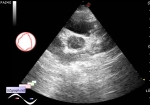
    On the current ultrasound: on the left side surface of the urine bladder(UB) outside is visualized anehogenous lesion up to 3x2x2 cm, at CFM without blood flow, in the lower part from the lesion in the direction of the wall of the UB is a tubular structure up to 5 mm in diameter (diverticulum of UB?).

    After the UB became empty: The lesion is visualized posteriorly from the UB and anterior to the uterus, the size of the lesion without significant dynamics.

    external link

:: attachments(2) ::

:: file 1 ::

:: file 2 ::


 Random 
Ultrasound cases - Testicular microlithiasis (microcalcification)


Ultrasound cases - Lateral neck cyst/ branchial cleft cyst
    

Ultrasound cases - Implanted metal plate
    

Ultrasound cases - PVL and criteria
    

Ultrasound cases - Where is the testicle?
    

 Dictionary 
LVEDD

PSV

LMP

IVC

Ved

LVEDV

LVESV

MC

LVOT

CFM


 Calculators 
TI-RADS

Qp:Qs

Teicholz systolic cardiac function

Body surface area

Pulmonary artery pressure


 Similar topics 
Ultrasound cases - Diffuse heterogeneous liver
    

Ultrasound cases - Postoperative abdominal wall fistula
    

Ultrasound cases - Stump appendicitis
    

Ultrasound cases - Liver scar after resection
    

Ultrasound cases - Subdiaphragmatic movable fluid collection (UFO)
    

Ultrasound cases - Lesion of the spleen or the case when it is easy to not notice
    

Ultrasound cases - I’m normal but others ...
    

Ultrasound cases - Single doubled kidney and Mullerian duct cyst in the infant
    

Ultrasound cases - Condition after removal of the spleen cyst
    

Ultrasound cases - Ileostoma stenosis and nephropathy