GB agenesis, Choledochal cysts, urolithiasis
Tags: Abdomen sonography, Urinary tract sonography, Gastrointestinal sonography, Female pelvis sonography, Images, Video, Clinical report, Esaote MyLab 70, Pediatric
| Posts | |||
| GB agenesis, Choledochal cysts, uro... | #1 |
| |||||
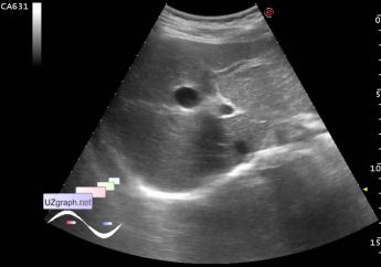
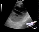
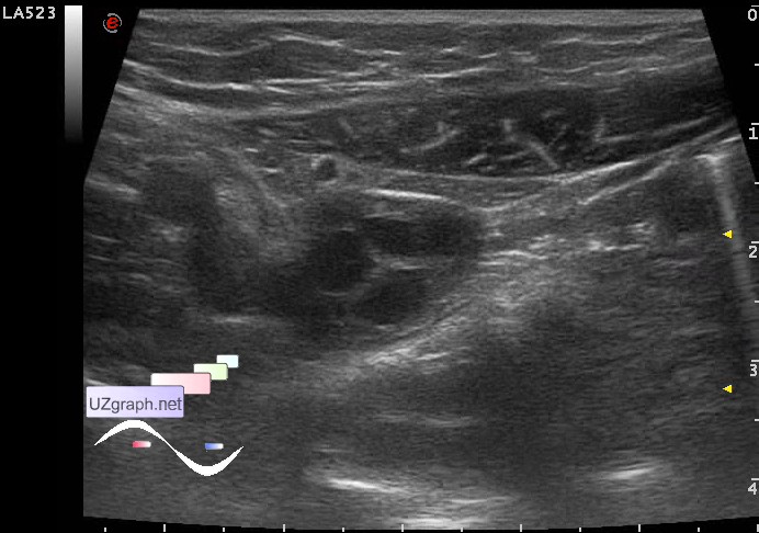
:: file 1 ::
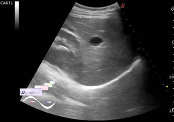
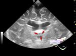
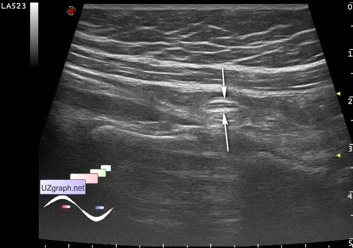
 :: file 2 ::
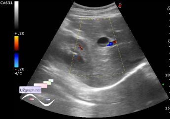
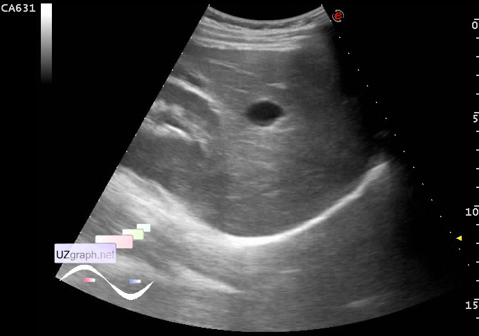 :: file 3 ::
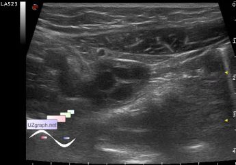
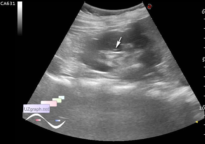
 :: file 4 ::
 :: file 5 ::
 :: file 6 ::
 :: file 7 ::
 :: file 8 :: | |||||