Log in    RU  


Main page :: Forum :: Ultrasound cases - Sonograms, cine-loops etc. ::

Parenchymal renal cyst


Tags: Urinary tract sonography, GE Vivid 3, Images, Video, Clinical report, Adult

   Posts
 
  Parenchymal renal cyst
#1

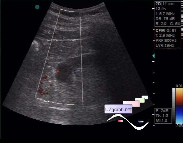
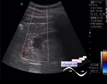
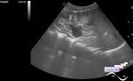
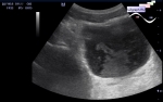
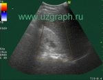
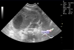
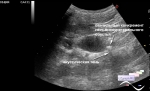
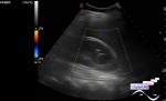
    The patient 78 years old came to the ultrasound of the kidneys, earlier by his words in one of the kidneys was found a cyst.

    On ultrasound in the projection of the parenchyma of the lower pole of the left kidney, a cyst is visualized up to 2 cm, at CFM a single signal of blood flow next to the cyst.

:: attachments(3) ::

:: file 1 ::

:: file 2 ::

:: file 3 ::


 Random 
Ultrasound cases - Combined (internal and external) hydrocephalus
    

Ultrasound cases - Acute cervical lymphadenitis
    

Ultrasound cases - Cyst but where?
    

Ultrasound cases - Condition after removal of the spleen cyst
    

Ultrasound cases - Ectopic thymus
    

 Dictionary 
IVSd

ESD

EFW

SF

FL

PVR

LAP

Freeze

BSA

LVEDD


 Calculators 
Body surface area

Myocardial mass

Type of an infant hip joint

Resistive index

Teicholz systolic cardiac function


 Similar topics 
Ultrasound cases - Autosomal recessive polycystic kidney disease (ARPKD)
    

Ultrasound cases - Bladder sludge
    

Ultrasound cases - Bright kidneys
    

Ultrasound cases - Ileostoma stenosis and nephropathy
    

Ultrasound cases - Renal pelvis clot
    

Ultrasound cases - Renal pelvis valve-stone
    

Ultrasound cases - Condition after treatment of a kidney cyst
    

Ultrasound cases - Hydronephrotic transformation & polypous bladder wall
    

Ultrasound cases - Pyelitis, cystitis
    

Ultrasound cases - Lesion in the projection of the adrenal gland 2