Log in    RU  


Main page :: Forum :: Ultrasound cases - Sonograms, cine-loops etc. ::

Lumbar pain


Tags: Urinary tract sonography, GE Vivid 3, Images, Video, Clinical report, Adult

   Posts
 
  Lumbar pain
#1

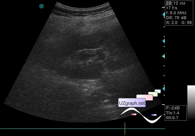
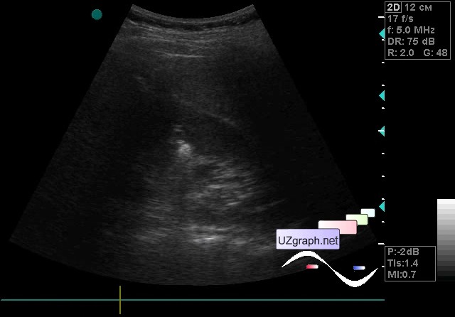
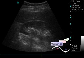
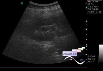
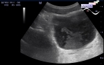
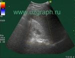
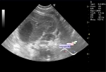
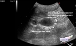
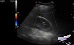
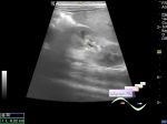
    A 27-year-old patient came to a kidney ultrasound with suspected urolithiasis, complaints of pain in the right lumbar region and frequent urination.

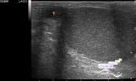
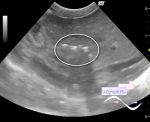
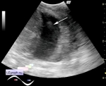
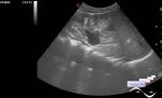
    On ultrasound in the right kidney multiple microliths and thickening of the renal pelvis wall up to 4 mm (pyelitis) are visualized, in the left kidney a single hyperechoic inclusion with acoustic shadow up to 6 mm is visualized(stone? calcificated papilla of the pyramid?).

    external link

:: attachments(4) ::

:: file 1 ::

:: file 2 ::

:: file 3 ::

:: file 4 ::


 Random 
Ultrasound cases - Thelarche
    

Ultrasound cases - Cubital vein thrombosis
    

Ultrasound cases - Intraventricular hemorrhage etc.
    

Ultrasound cases - Intestinal intussusception, intestinal obstruction, ascites ...
    

Ultrasound cases - Richly vascularized thyroid nodule
    

 Dictionary 
EDV

TV

CO

ESV

PA

Anatomical M-mode

EDD

RVOT

Ved

ESD


 Calculators 
TI-RADS

Type of an infant hip joint

Teicholz systolic cardiac function

Thyroid volume

Gestational age by Ultrasound


 Similar topics 
Ultrasound cases - Autosomal recessive polycystic kidney disease (ARPKD)
    

Ultrasound cases - Bladder sludge
    

Ultrasound cases - Bright kidneys
    

Ultrasound cases - Ileostoma stenosis and nephropathy
    

Ultrasound cases - Renal pelvis clot
    

Ultrasound cases - Renal pelvis valve-stone
    

Ultrasound cases - Condition after treatment of a kidney cyst
    

Ultrasound cases - Hydronephrotic transformation & polypous bladder wall
    

Ultrasound cases - Pyelitis, cystitis
    

Ultrasound cases - Lesion in the projection of the adrenal gland 2