Log in    RU  


Main page :: Forum :: Ultrasound cases - Sonograms, cine-loops etc. ::

Birads 5


Tags: Breast sonography, GE Vivid 3, Images, Video, Clinical report, Adult

   Posts
 
  Birads 5
#1

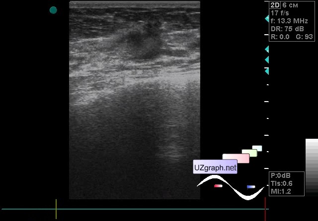
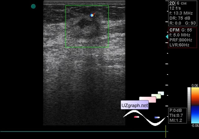
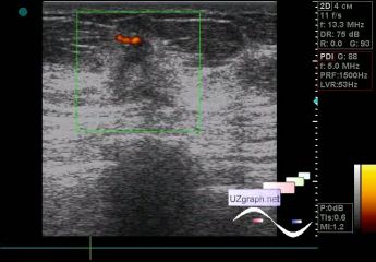
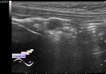
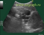
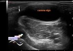
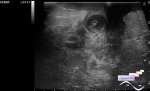
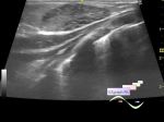
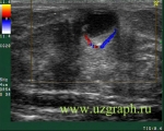
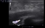
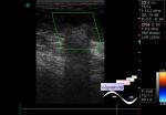
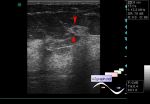
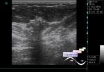
    A 51-year-old woman, without complaints, came to a screening ultrasound of the mammary glands; earlier, according to her, was found an adenoma in another mammary gland at mammography, which was not seen later on an ultrasound.

    On the current ultrasound in the other mammary gland, next to the nipple, mainly hypoechoic mass of a bizarre form, with a fuzzy uneven contour, acoustic shadow, vertical orientation, radiant anterior edge, blood flow is mapped at the CFM and PD in this radiant edge (birads 5, supposedly malignant ).

    The mass is dense - when a probe passes over it, the probe jumps over it.

    Regional LNs are not visualized.

    Urgent consultation of the mammologist and puncture was recommended.

    In the coronary view(see attachment file 5), we see a characteristic view of the " sun" or " octopus" , i.e. deformation of surrounding tissues.

    external link

:: attachments(5) ::

:: file 1 ::

:: file 2 ::

:: file 3 ::

:: file 4 ::

:: file 5 ::

  13:22 15-12-2018
 
#2

    The management reported that the cancer was verified.


 Random 
Ultrasound cases - Large axillary lesion
    

Ultrasound cases - Cryptorchidism, microlithiasis, bladder lesion
    

Ultrasound cases - Meconium peritonitis and ascites
    

Ultrasound cases - Extreme spleen
    

Ultrasound cases - Actualization of the ASA
    

 Dictionary 
RAP

Gain

ESV

GA

LMP

Aliasing doppler artifact

Freeze

HR

IVC

PASP


 Calculators 
Resistive index

Body surface area

Type of an infant hip joint

Pulmonary artery pressure

TI-RADS


 Similar topics 
Ultrasound cases - Mastitis and cysts
    

Ultrasound cases - Neonatal mastitis
    

Ultrasound cases - Adolescent Gynecomastia
    

Ultrasound cases - Subacute mastitis (cyst with inflammation) in a teenager
    

Ultrasound cases - Breast Cysts
    

Ultrasound cases - Infant isolated thelarche
    

Ultrasound cases - Another Birads 4-5
    

Ultrasound cases - Star-like lesion
    

Ultrasound cases - Birads 4-5
    

Ultrasound cases - Breast lesions 3