Log in    RU  


Main page :: Forum :: Ultrasound cases - Sonograms, cine-loops etc. ::

PVL, pericarditis, LV fibroelastosis, ascites


Tags: Brain sonography(Neurosonography), Cardiac sonography(Echocardiography), Abdomen sonography, GE Logiq Book XP, Images, Video, Clinical report, Pediatric

   Posts
 
  PVL, pericarditis, LV fibroelastosi...
#1

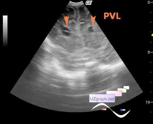
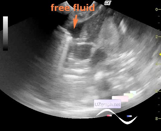
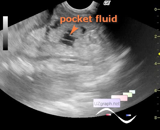
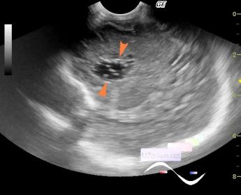
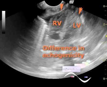
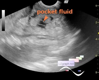
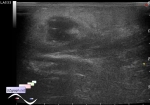
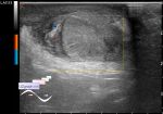
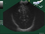
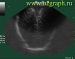
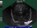
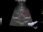
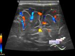
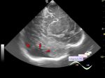
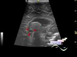
    A newborn in the reanimation unit with PVL (periventricular leukomalacia) and polyserositis, ultrasound for fluid was prescribed.

    On ultrasound, there is a pocket fluid in the abdominal cavity, also a little amount of fluid, most likely in the pericardial cavity, near the right heart ventricle (pericarditis?) with visual signs of compression, and then something heterogeneous in the region of the apex of the heart in oblique view, like a pocket fluid, was evident, nevertheless, most likely this is the wall of the right ventricle, if anyone noticed, the left ventricular myocardium looks more echogenic (endocardial LV fibroelastosis?), thats why such visualization present.

    external link

:: attachments(6) ::

:: file 1 ::

:: file 2 ::

:: file 3 ::

:: file 4 ::

:: file 5 ::

:: file 6 ::


 Random 
Ultrasound cases - Pain in groin
    

Ultrasound cases - Cyst of the epididymis and appendages
    

On-call room - Orange worm in medical equipment

Ultrasound cases - Neonatal testicular torsion
    

 Dictionary 
TVUS

Aliasing doppler artifact

LVESV

M-mode

LVOT

Doppler angle

VTI

PSV

IVC

CO


 Calculators 
Thyroid volume

Gestational age by Ultrasound

Myocardial mass

Type of an infant hip joint

Resistive index


 Similar topics 
Ultrasound cases - Echogenic brain foci
    

Ultrasound cases - Subdural hemorrhage 2
    

Ultrasound cases - Willis circle at ultrasound
    

Ultrasound cases - Periventricular hyperechogenicity
    

Ultrasound cases - Combined (internal and external) hydrocephalus
    

Ultrasound cases - Subdural hemorrhage
    

Ultrasound cases - Cyst in the projection near the posterior horn of the lateral ventricle
    

Ultrasound cases - Subdural collection
    

Ultrasound cases - Anechogenic inclusion in the posterior cranial fossa
    

Ultrasound cases - Cyst of the base of the brain