Log in    RU  


Main page :: Forum :: Ultrasound cases - Sonograms, cine-loops etc. ::

"Ball" in the abdomen


Tags: Soft tissues sonography, Esaote MyLab 70, Clinical report, Pediatric

   Posts
 
  "Ball" in the abdomen
#1

    A 10-year-old child with abdominal pain syndrome was admitted to the emergency department of the Children's City Clinical Hospital, and was urgently sent for an ultrasound scan.

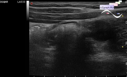
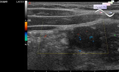
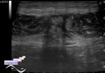
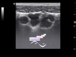
    On ultrasound in the right mesogastrium, multiple enlarged mesenteric lymph nodes are visualized (differential diagnosis: mesadenitis, etc.), next to them a hypoechoic lesion of a rounded shape is visualized, without echo-pattern of the lymph node echogenic hilus, with increased blood flow at CFM and atypical vascular pathways - peripherally diametrically located CFM signals, horseshoe-like (risk of mts 60%), suspicious of neoangiogenesis (differential diagnosis: mts, etc.).

    Consultation of the oncologist was recommended.


 Random 
Ultrasound cases - Postoperative abdominal wall fistula
    

Ultrasound cases - Curved small popliteal cyst
    

Ultrasound cases - Accessory spleen
    

Ultrasound cases - Subdural collection
    

Ultrasound cases - Intestine contusion
    

 Dictionary 
PA

HC

CO

MC

B-mode

M-mode

GA

CW

LVESD

TVUS


 Calculators 
Teicholz systolic cardiac function

Volume (ellipse formula)

Pressure gradient

Type of an infant hip joint

Pulmonary artery pressure


 Similar topics 
Ultrasound cases - Penetrating injury of the femur
    

Ultrasound cases - Cephalohematoma
    

Ultrasound cases - Urachus fistula
    

Ultrasound cases - Cranial mass
    

Ultrasound cases - Pilonidal coccyx cyst
    

Ultrasound cases - Palm fistula, in search of pencil lead
    

Ultrasound cases - Postoperative abdominal wall fistula
    

Ultrasound cases - Nucks cyst
    

Ultrasound cases - Paraproctitis
    

Ultrasound cases - Submandibular area lesion